3-d ultrasound imaging

a technology of ultrasound imaging and reference view, applied in the field of ultrasound imaging system, can solve the problems of time-consuming and laborious, deviating from the standard view that allows reliable diagnosis, and unable to obtain a reference view that is sufficiently good, so as to achieve quick and reliable analysis of volume data.

Inactive Publication Date: 2011-08-18
KONINKLIJKE PHILIPS ELECTRONICS NV
View PDF6 Cites 49 Cited by
  • Summary
  • Abstract
  • Description
  • Claims
  • Application Information

AI Technical Summary

Benefits of technology

There is a need for an improved ultrasound imaging system, which allows a quicker and more reliable analysis of volume data.
The anatomy-related description can be used to advantage in processing steps that are applied to the volume data for the purpose of analyzing this data and establishing a diagnosis. The anatomy-related description may guide, as it were, such processing steps. Identifying anatomic features is no longer a process that substantially depends on operator abilities and that requires relatively sophisticated skills. Standard views that are obtained from the volume data will be less operator dependent, and can be obtained with less skill required on the part of the operator, which allows a more reliable diagnosis. For example, in obstetric applications, this will typically result in a higher probability of detecting structural anomalies in a fetal heart. There is less risk that such anomalies remain undetected. Moreover, the anatomy-related description allows speeding up a process of manipulating and analyzing the volume data.
The ultrasound imaging system preferably comprises a user interface that allows an operator to define a minimization criterion that is to be used in finding the best match between the volume data and the geometrical model.

Problems solved by technology

The method described in the aforementioned patent application suffers from several limitations.
In case the reference view is imprecise, views that are obtained on the basis of the spatial mathematical relationships will also be imprecise and thus deviate from standard views that allow reliable diagnosis.
Moreover, obtaining a reference view that is sufficiently good can be time consuming, in particular for a less experienced operator.
These manipulations are also subject to possible errors and can be time consuming too.
However, there may be significant variability in terms of geometric properties of the organ concerned, even in normal cases.
However, this may be confusing for the operator, and may therefore lead to errors.
Moreover, such an approach is rather inefficient.

Method used


Image

  • 3-d ultrasound imaging
  • 3-d ultrasound imaging
  • 3-d ultrasound imaging

Examples


Embodiment Construction

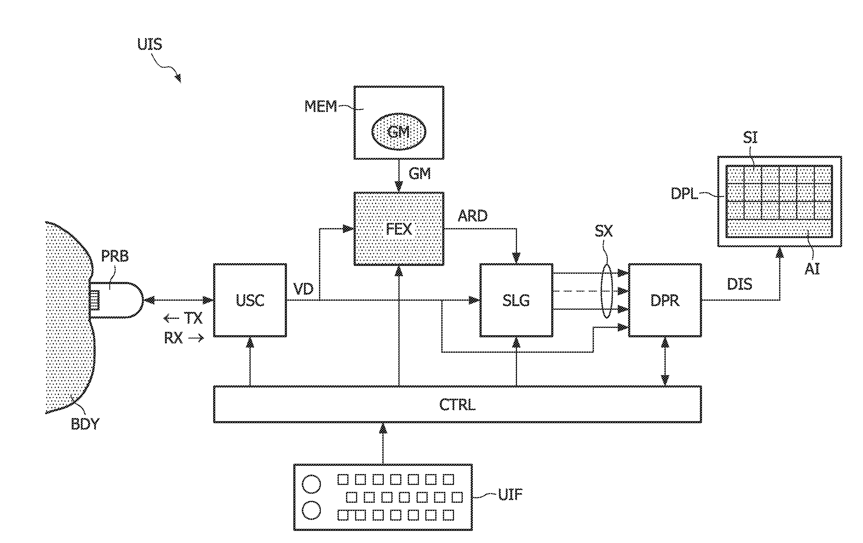
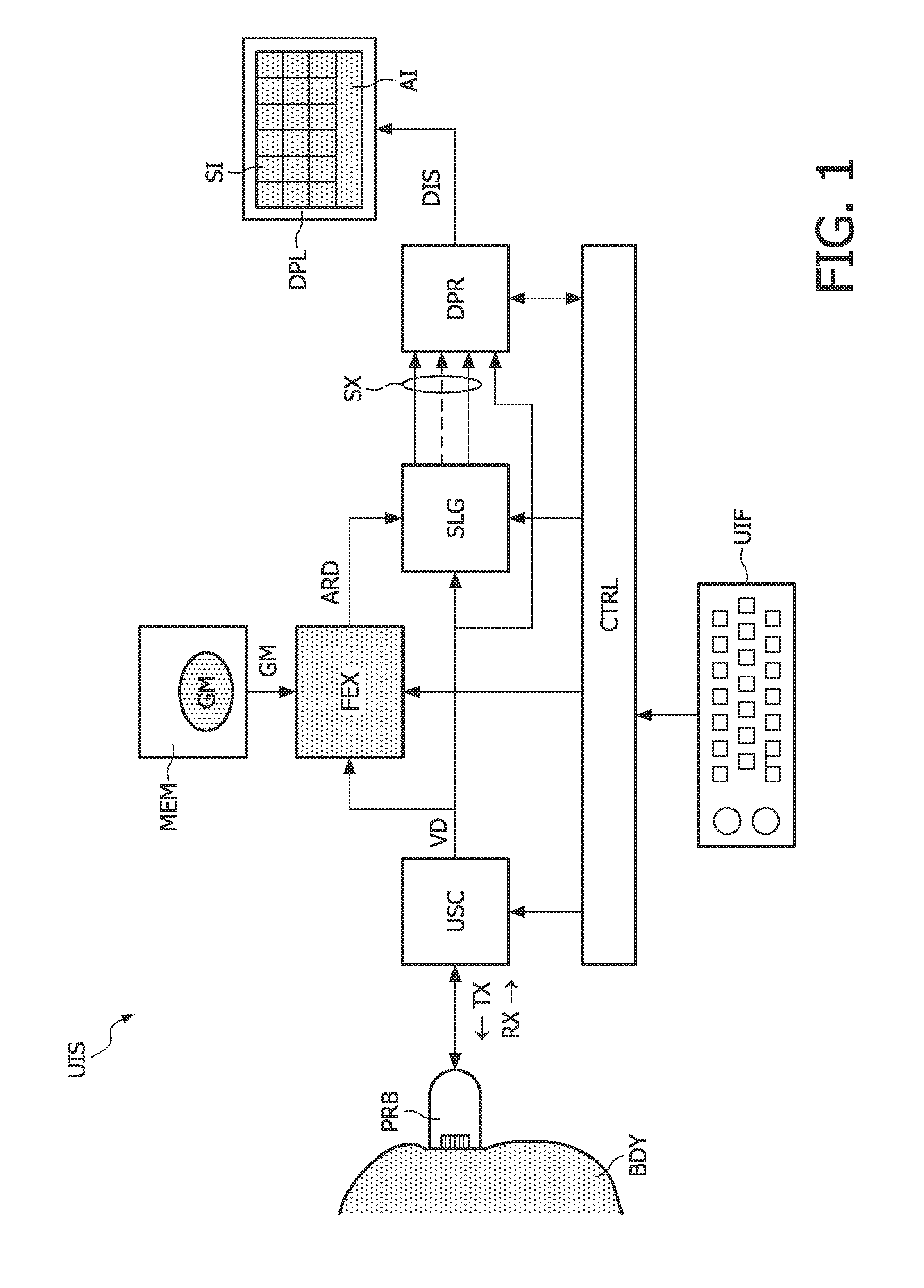
FIG. 1 illustrates an ultrasound imaging system UIS, which capable of carrying out a 3-D ultrasound scan. The ultrasound imaging system UIS comprises various functional entities that constitute an ultrasound imaging acquisition-and-processing path: a probe PRB, an ultrasound scanning assembly USC, a feature extractor FEX, a slice generator SLG, and a display processor DPR. The probe PRB may comprise, for example, a two-dimensional array of piezoelectric transducers. The ultrasound scanning assembly USC may comprise an ultrasound transmitter and an ultrasound receiver, which may each include a beam-forming module. The ultrasound scanning assembly USC may further comprise one or more filter modules, a so-called B-mode processing module, and a Doppler-mode processing module.

The feature extractor FEX may be implemented by means of, for example, a set of instructions that has been loaded into a programmable processor. In such a software-based implementation, the set of instructions defin...


PUM


Abstract

In an ultrasound imaging system (UIS), an ultrasound scanning assembly (USC) provides volume data (VD) resulting from a three-dimensional scan of a body (BDY). A feature extractor (FEX) searches for a best match between the volume data (VD) and a geometrical model (GM) of an anatomical entity. The geometrical model (GM) comprises respective segments representing respective anatomic features. Accordingly, the feature extractor (FEX) provides an anatomy-related description (ARD) of the volume data (VD), which identifies respective geometrical locations of respective anatomic features in the volume data (VD). In a preferred embodiment, a slice generator (SLG) generates slices (SX) from the volume data (VD) based on the anatomy-related description (ARD) of the volume data (VD).

Description

FIELD OF THE INVENTIONAn aspect of the invention relates to an ultrasound imaging system that is capable of carrying out a three-dimensional (3-D) ultrasound scan, which produces volume data. The ultrasound imaging system may be helpful in, for example, fetal examinations, in particular detection of structural anomalies in a fetal heart. Other aspects of the invention relate to a method of ultrasound imaging, and a computer program product.BACKGROUND OF THE INVENTIONA 3-D ultrasound scan typically involves emitting ultrasound waves that illuminate, as it were, a particular volume within a body, which may be designated as target volume. This can be achieved, for example, by emitting ultrasound waves at multiple different angles. Volume data is obtained by receiving and processing reflected waves. The volume data is a representation of the target volume within the body. The volume data can be displayed on a display device in a fashion that provides a three-dimensional representation, ...

Claims


Application Information

Patent Timeline
Patent Type & Authority Applications(United States)
IPC IPC(8): A61B8/14
CPCA61B8/0833A61B8/0866A61B8/0883A61B8/523A61B8/5238G01S15/8977G01S15/8993A61B8/483
Inventor COLLET-BILLON, ANTOINEJAGO, JAMESDOW, ALASDAIRPUMPHREY, LISA
Owner KONINKLIJKE PHILIPS ELECTRONICS NV
Why Eureka
Try Eureka