Method and system for detecting lung tumors and nodules

Inactive Publication Date: 2011-10-20
UNIVERSITY OF ROCHESTER
View PDF32 Cites 35 Cited by
  • Summary
  • Abstract
  • Description
  • Claims
  • Application Information

AI Technical Summary

Benefits of technology

[0012]The aim of the present invention is to demonstrate a novel, fully automatic computer detection method applicable to metastatic tumors to the lung with a diameter of 4-20 mm in high-risk patients using typical computed tomography (CT) scans of the chest. In the present invention, three-dimensional (3D) spherical tumor appearance models (templates) of various sizes were created to match representative CT imaging parameters and to incorporate partial volume effects. Taking into account the variability in the location of CT sampling planes cut through the spherical models, three offsetting template models were created for each appearance model size. Lung volumes were automatically extracted from computed tomography images and the correlation coefficients between the sub-regions around each voxel in the lung volume and the set of appearance models were calculated using a fast frequency domain algorithm. To determine optimal parameters for the templates, simulated tumors of varying sizes and eccentricities were generated and superposed onto a representative human chest image dataset. The method was applied to real image sets from twelve patients with kno

Problems solved by technology

Unfortunately, traditionally the utility of radiation to control lung disease has been limited by the lungs' poor radio-tolerance.
At small sizes, however, the presence of metastatic tumors is not discerned by the patient and not detectable using clinical pulmonary function tests, hence the need for lung tumor screening via medical imaging.
To read and interpret these massive amounts of image data requires substantial amount of radiologist effort and predisposes the screening process to human error and missed detection of cancerous lesions.
Results from these and related CAD algorithms are encouraging in general; however, most current CAD schemas suffer from a miss-rate of 10-30% (low sensitivity) and, at the same time, generate a large number of false-positives (low specificity).
A high false positive rate is undesirab

Method used


Image

  • Method and system for detecting lung tumors and nodules
  • Method and system for detecting lung tumors and nodules
  • Method and system for detecting lung tumors and nodules

Examples


Embodiment Construction

[0026]Hereinafter, exemplary embodiments of the present invention will be described in detail. However, the present invention is not limited to the embodiments disclosed below, but can be implemented in various forms. The following embodiments are described in order for this disclosure to be complete and enabling of practice of the invention by those of ordinary skill in the art.

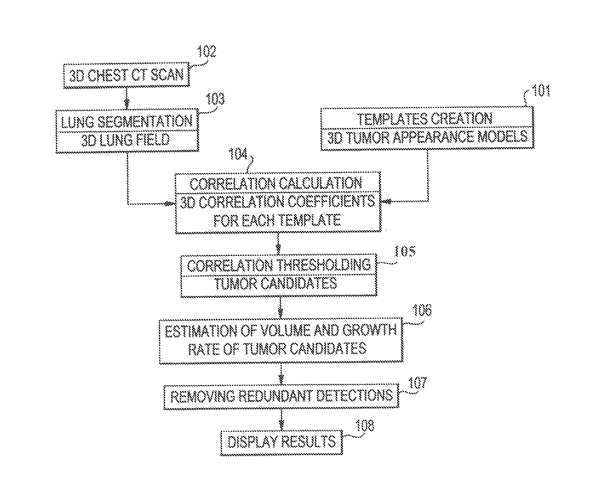
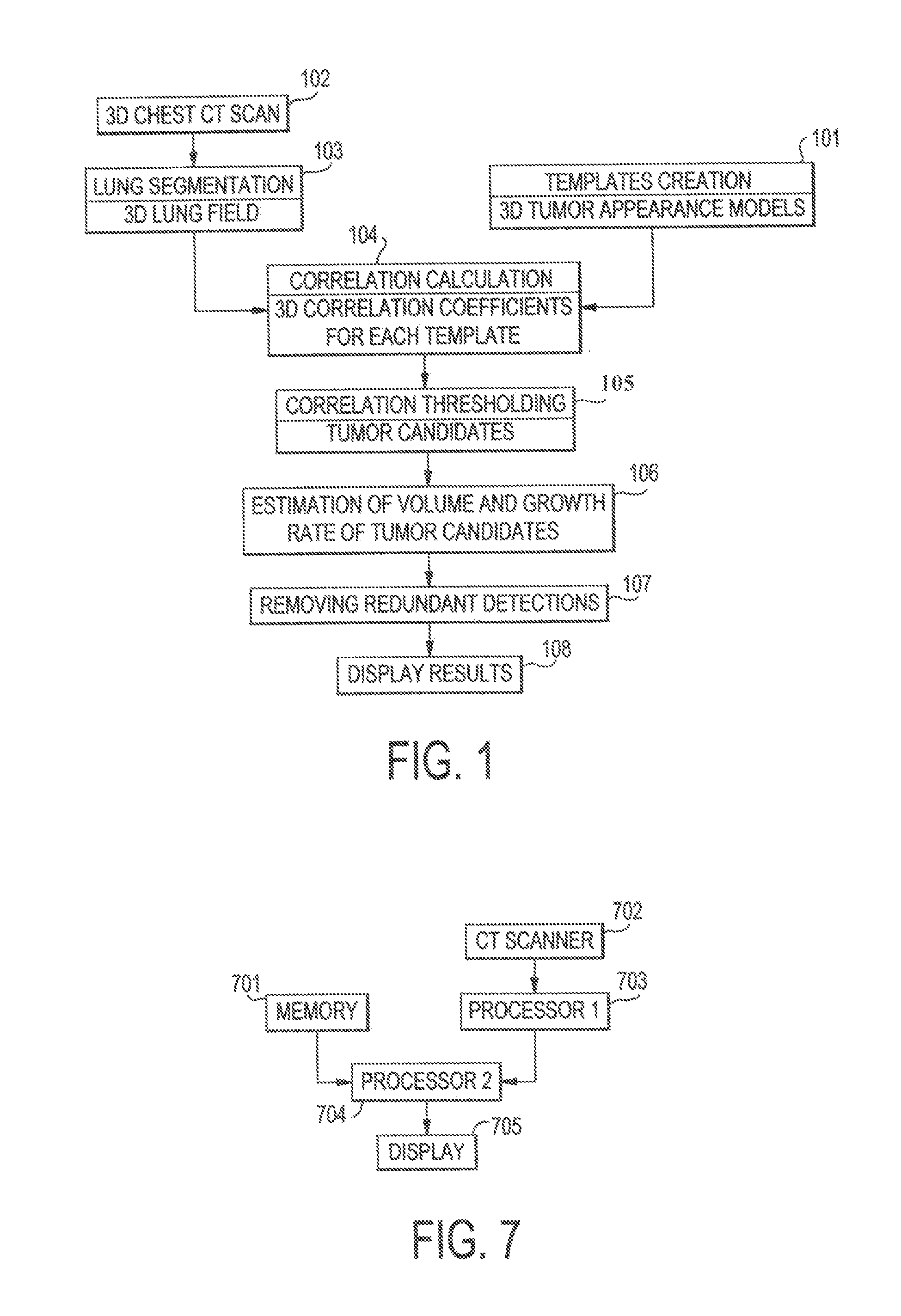
[0027]FIG. 1 shows a flowchart of a process of a system for detecting lung tumors or nodules according to one exemplary embodiment of the present invention. In step 101, templates of 3D appearance models of tumors are created. In step 102, 3D CT scans of the chest of a patient are obtained. The scans show images of slices of the chest. In step 103, lung segmentation is processed. As described in detail below, this process produces 3D imaging data of the lung parenchyma without the surrounding soft tissue or bones and without the blood vessels, lesions, or the like inside the lung region. The 3D imaging data ...


PUM


Abstract

A method and system for detecting tumors and nodules in a lung tissue are provided. The method includes the steps of (a) providing a plurality of asymmetric templates of at least one 3D appearance model of a nodule; (b) providing a 3D imaging data set of the entire area of a tissue; (c) matching the 3D imaging data set of the entire area of the tissue with each of the plurality of asymmetric templates to search for 3D objects in the tissue that match the at least one 3D appearance model; (d) determining the volume of the 3D objects found in the tissue; and (e) providing an output representing 3D objects that match the 3D appearance model.

Description

CROSS-REFERENCE TO RELATED APPLICATIONS[0001]This application is the U.S. national phase of PCT International Patent Application No. PCT / US2008 / 068407, filed Jun. 26, 2008, published on Dec. 31, 2008, as WO 2009 / 003128 A2, which claims the benefit of U.S. Provisional Patent Application No. 60 / 929,421, filed Jun. 26, 2007 in the U.S. Patent and Trademark Office, titled “IMAGE PROCESSING METHOD FOR COMPUTING VOLUME OF TUMORS AND NODULES FROM ED MEDICAL IMAGES”, the entire disclosure of which is incorporated herein by reference.[0002]This application is also related to the subject matter disclosed in U.S. Provisional Patent Application No. 60 / 598,844, filed Aug. 5, 2004 in the U.S. Patent and Trademark Office, titled “AUTOMATIC COMPUTER AIDED DETECTION OF TUMOR USING 3D TEMPLATE MATCH,” the entire disclosure of which is incorporated herein by reference.FIELD OF THE INVENTION[0003]The present invention relates to a method and device for detecting tumors in a lung tissue, and more partic...

Claims


Application Information

Patent Timeline
IPC IPC(8): G06K9/00
CPCG06T7/0014G06T2207/30064G06T2207/10081G06T7/401G06T7/41
Inventor O'DELL, WALTERAMBROSINI, ROBERTWANG, PENG
Owner UNIVERSITY OF ROCHESTER
Why Eureka
Try Eureka