System and method for image-guided arthroscopy

Inactive Publication Date: 2011-11-03
REPRISE TECH
View PDF4 Cites 27 Cited by
  • Summary
  • Abstract
  • Description
  • Claims
  • Application Information

AI Technical Summary

Problems solved by technology

One of the challenges with camera technologies is that they are not very well suited for imaging through blood, fascia, or other tissues which may not transmit light as well as saline, synovial fluid, or gases such as nitrogen, carbon dioxide, or air.
For example, arthroscopy of the hip presents the clinician with various challenges, including the desire to have minimal port wounds and port sizes, as well as minimal tissue da

Method used


Image

  • System and method for image-guided arthroscopy
  • System and method for image-guided arthroscopy
  • System and method for image-guided arthroscopy

Examples


Embodiment Construction

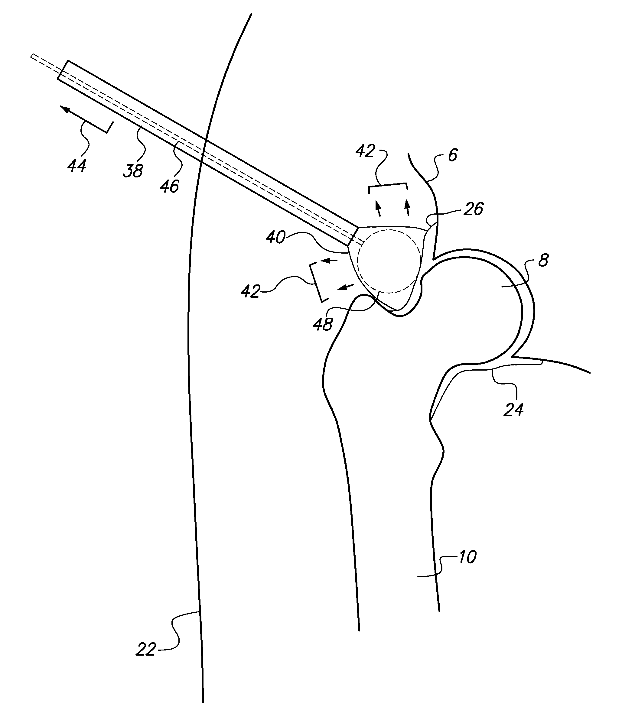
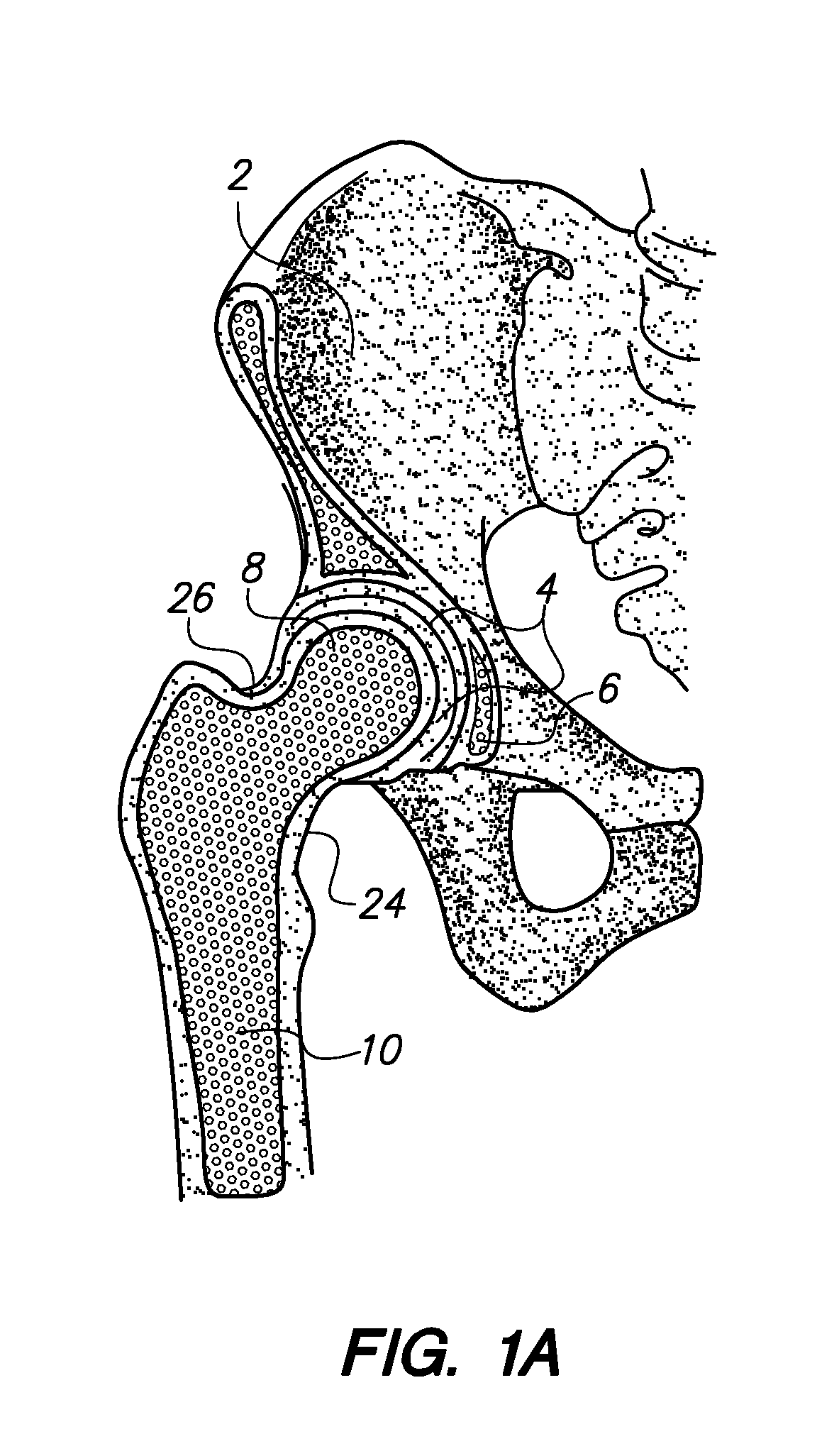
[0013]Referring to FIG. 2A, at the extremes of joint mobility, the capsule (24, 26) may become strained, and the geometry of the calcified structures, such as the acetabulum (6) and femur (8), may ultimately cause such structures to become mechanical movement limits. Referring to FIGS. 2B and 2C, certain patients may impinge soft tissue structures as well in such extreme joint positions, such as the labrum (28) around the joint socket formed by the three bones of the acetabulum (6) when loaded against a portion of the neck of the femur (8). Removal of portions (30) of the femoral neck calcified tissue may assist in reducing this type of intersection, as described, for example, in U.S. Patent Application Ser. No. 61 / 305,519, which is incorporated by reference herein in its entirety. Notwithstanding such procedures, it is desirable in many interventional scenarios to address damaged labral tissue problems, and also address bone geometry configurations which can be optimized with minim...


PUM


Abstract

Configurations are described for conducting minimally invasive medical diagnoses and interventions utilizing elongate instruments and assemblies comprising one or more imaging devices, and one or more remote retraction and distraction devices. Retraction and distraction devices, such as balloons, mechanical retraction members, and/or trocar screw geometries may be utilized to access, investigate, and intervene at the joint capsule, or inside of the joint capsule. Imaging devices, such as optical image capture devices, ultrasound transducers, and optical coherence tomography fibers, may be utilized to assist with navigation of the pertinent tools during diagnostic and interventional steps.

Description

RELATED APPLICATION DATA[0001]The present application claims the benefit under 35 U.S.C. §119 to U.S. provisional patent application Ser. Nos. 61 / 305,519, filed Feb. 17, 2010, 61 / 311,117, filed Mar. 5, 2010, 61 / 315,795, filed Mar. 19, 2010, and 61 / 377,670, filed Aug. 27, 2010. The foregoing applications are hereby incorporated by reference into the present application in their entirety.FIELD OF THE INVENTION[0002]The present invention relates to diagnostic and / or interventional arthroscopy, and particularly to the utilization of various imaging, retraction, and distraction technologies as coupled to elongate arthroscopy instruments for improved visualization, navigation, access, and guidance during clinical procedures.BACKGROUND[0003]Hundreds of thousands of patients in the US and other countries undergo diagnostic and / or interventional arthroscopy every year. For example, arthroscopy of the knee joint, hip joint, and elbow joint is relatively common, and generally is associated wit...

Claims


Application Information

Patent Timeline
IPC IPC(8): A61M29/00A61B1/04
CPCA61B1/313A61B1/32A61B17/0218A61B17/025A61B17/885A61B17/3439A61B17/3472A61B2017/0268A61B2017/320048A61B17/34A61B17/3423A61B2017/0225A61B2017/3486
Inventor LITVACK, FRANKKLAPPER, ROBERT C.SHANLEY, JOHN F.
Owner REPRISE TECH
Why Eureka
Try Eureka