Interventions Using Correlated Nuclear and Ultrasound Imaging

a nuclear and ultrasound technology, applied in the field of medical imaging, can solve the problems of guiding interventions using these methods, raylman fails to teach apparatus or methods to perform procedures within clinically acceptable time limits, and pelizzari fails to disclose apparatus or methods to utilize precise correlative methods, etc., to achieve the effect of quick and accurate positioning of interventional devices and simplified task of interventional device localization

Inactive Publication Date: 2011-12-01
YARNALL STEPHEN T
View PDF4 Cites 6 Cited by
  • Summary
  • Abstract
  • Description
  • Claims
  • Application Information

AI Technical Summary

Benefits of technology

[0012]In one aspect of the present invention an apparatus and method is provided that can be utilized to quickly and accurately position an interventional device in relation to a lesion of interest by first identifying the location of the lesion in two dimensions using nuclear emission imaging and then by determining the third dimension using ultrasound imaging applied precisely at the two-dimens...

Problems solved by technology

Stereotactic methods using nuclear-emission imaging have been developed to position a device in relation to a lesion for performing an intervention; however, guiding interventions using these methods can be problematic due to procedure time constraints, lesion movement, interventional device control, and the three-dimensional resolving power of the nuclear image.
However, Pelizzari fails to disclose an apparatus or method means to utilize precise correlative methods without additional ionizing radiation.
However, Raylman fails to teach apparatus or method to perform the procedure within clinically-acceptable time limits, to accommodate necessary redirection of interventional device during insertion due to tissue characteristics or mis-targeting, and to confirm the actual location of the interventional device with respect to the lesion.
However, Ashburn fails to teach an apparatus or method to perform the procedure within clinically-acceptable time limits, to accommodate necessary redirection of interventional device during insertion due...

Method used


Image

  • Interventions Using Correlated Nuclear and Ultrasound Imaging
  • Interventions Using Correlated Nuclear and Ultrasound Imaging
  • Interventions Using Correlated Nuclear and Ultrasound Imaging

Examples


Embodiment Construction

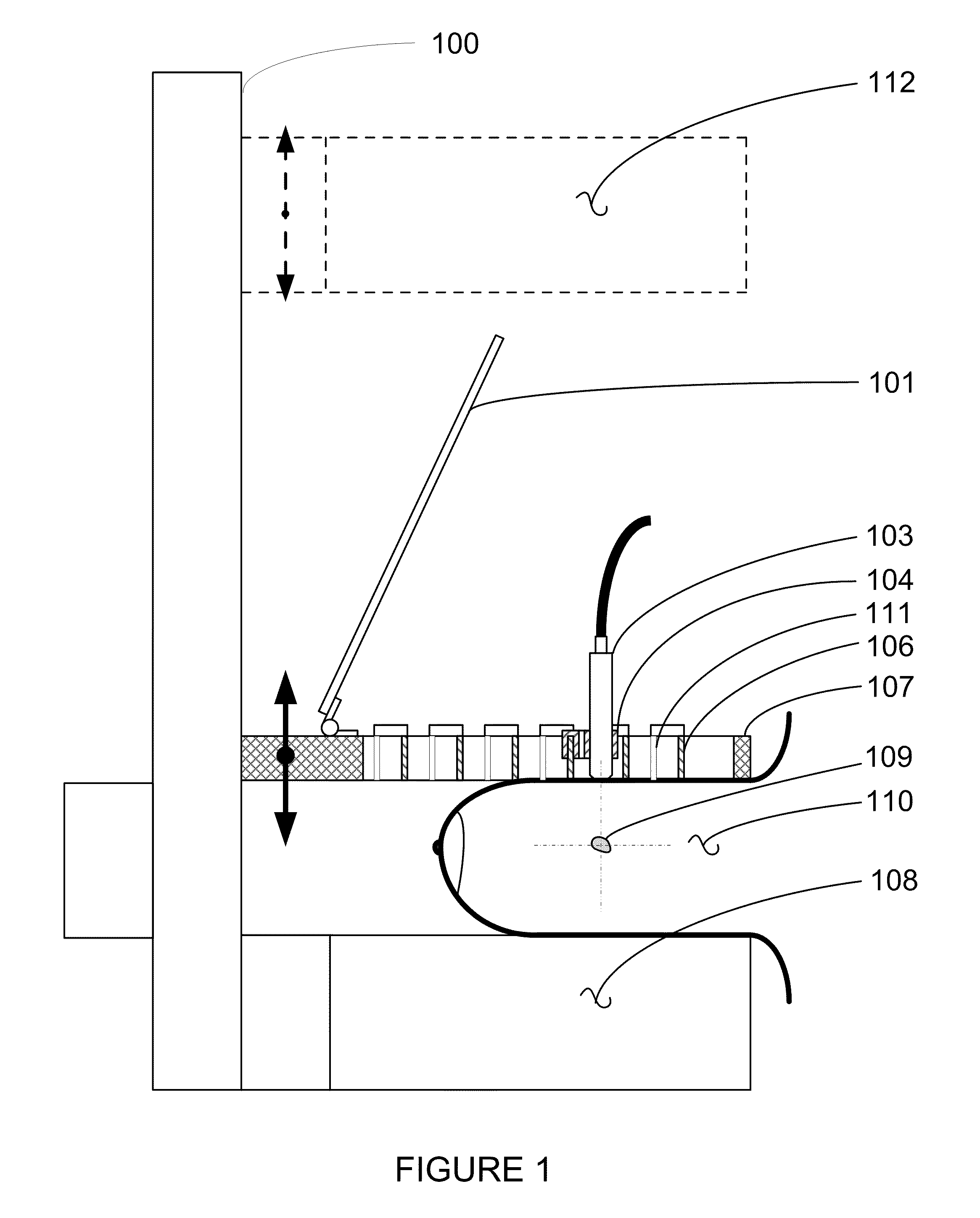
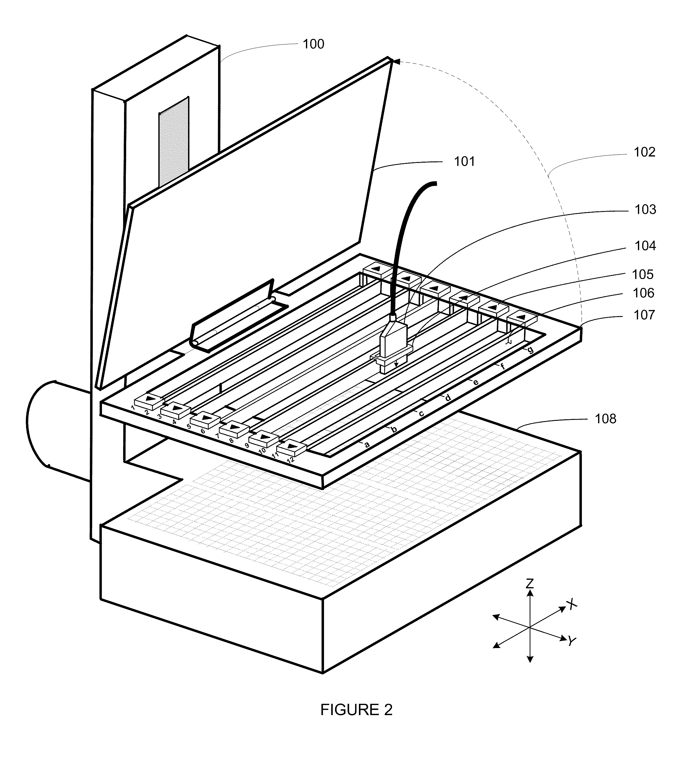
[0015]Nuclear-emission imaging is sometimes used to identify abnormal cellular function(s) (e.g. glucose metabolism as with FDG-PET, mitochondrial proliferation as with sestamibi-scintigraphy) of a region of tissue (lesion) that may be associated with tumor growth (neoplasia). However, it is difficult to use nuclear-emission imaging alone to guide interventions due to the long image acquisition periods (e.g. five minutes), unacceptable nuclear-emission image spatial resolution and the transparency of most interventional devices to emission imaging.

[0016]Ultrasound imaging may be utilized to identify very subtle abnormalities in tissue density and structure that commonly result from neoplasia. However, abnormalities produced by cancer are difficult to distinguish from abnormalities produced by benign processes using ultrasound, and ultrasound alone is considered to have unacceptable specificity for early breast cancer detection. Notwithstanding, it is commonly understood that most su...


PUM


Abstract

An apparatus and method for localizing a nuclear-emitting lesion during an intervention using correlated nuclear and ultrasound imaging. The apparatus provides interventional access and quasi-stereotactic positioning of interventional devices, with real-time ultrasound image visualization for tracking the approach of the device to the lesion. The apparatus is intended to overcome the shortcomings of fully-stereotactic nuclear-emission image localization.

Description

CROSS-REFERENCE TO RELATED APPLICATION[0001]The present application claims the benefit under 35 U.S.C. §119(e) of U.S. Provisional Application No. 61 / 348,730, filed May 26, 2010, which is incorporated herein by reference.FIELD OF THE INVENTION[0002]The present invention relates generally to the field of medical imaging. More specifically, the invention relates to systems and methods for using nuclear-emission imaging to localize lesions for tissue harvesting or marking.BACKGROUND OF THE INVENTION[0003]In medicine, nuclear-emission imaging is commonly utilized to identify regions (lesions) that may contain abnormal cells, and must be sampled, marked or otherwise treated using interventional devices. Stereotactic methods using nuclear-emission imaging have been developed to position a device in relation to a lesion for performing an intervention; however, guiding interventions using these methods can be problematic due to procedure time constraints, lesion movement, interventional dev...

Claims


Application Information

Patent Timeline
IPC IPC(8): A61B6/00A61B8/00
CPCA61B6/037A61B6/0414A61B6/12A61B6/481A61B6/502A61B6/4417A61B8/0825A61B8/0841A61B8/4416A61B8/5238A61B6/5247
Inventor YARNALL, STEPHEN T.
Owner YARNALL STEPHEN T
Why Eureka
Try Eureka