Dual screen radiographic detector with improved spatial sampling

a radiographic detector and spatial sampling technology, applied in the field of digital radiographic detectors, can solve the problems of double exposure technique sensitive to patient motion artifacts, misregistration between, and loss of spatial resolution of medical x-ray detectors employing scintillating phosphor screens to absorb x-rays and produce light,

Active Publication Date: 2011-12-15
CARESTREAM HEALTH INC
View PDF7 Cites 52 Cited by
  • Summary
  • Abstract
  • Description
  • Claims
  • Application Information

AI Technical Summary

Benefits of technology

[0014]Embodiments according to the present invention address the foregoing needs in the art by providing an improved radiographic imaging apparatus having a first scintillator, a second scintillator, a plurality of first photosensitive elements, and a plurality of second photosensitive elements. The first scintillator has first scintillator properties and the second scintillator has second scintillator properties different from the first scintillator properties. The plurality

Problems solved by technology

Generally, medical X-ray detectors employing a scintillating phosphor screen to absorb X-rays and produce light suffer the loss of spatial resolution due to lateral light diffusion in the phosphor screen.
Since a double exposure of the patient must be performed, and the switching of the X-ray tube voltage must take a finite time, the double exposure technique would be sensitive to patient motion artifacts and to misregistration between the two images.
While digital

Method used


Image

  • Dual screen radiographic detector with improved spatial sampling
  • Dual screen radiographic detector with improved spatial sampling
  • Dual screen radiographic detector with improved spatial sampling

Examples


Embodiment Construction

[0029]Embodiments according to the application will be described with reference to the accompanying figures.

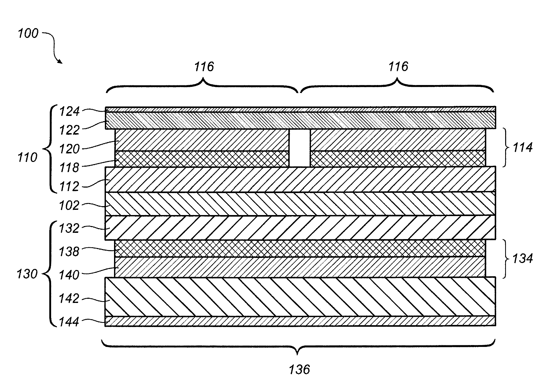
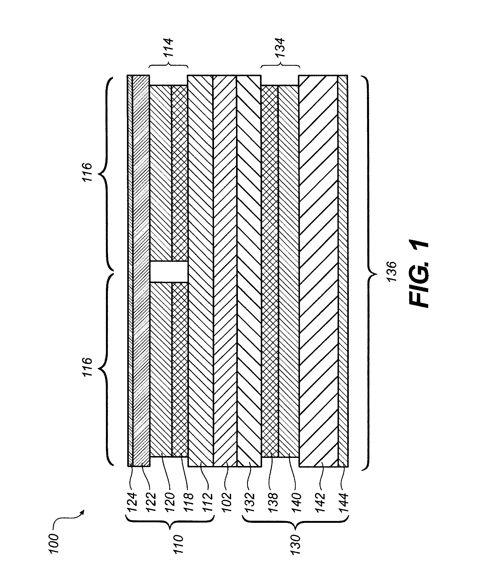
[0030]FIG. 1 illustrates a first embodiment of a dual screen radiographic imaging apparatus 100. Generally speaking, the dual screen radiographic imaging apparatus 100 includes a first detector 110 and a second detector 130. In the example of FIG. 1, the first detector 110 and the second detector 130 are separated by and disposed on the opposite sides of a filter 102. According to its use thereof, the filter 102 can be an x-ray filter and / or an optical filter.

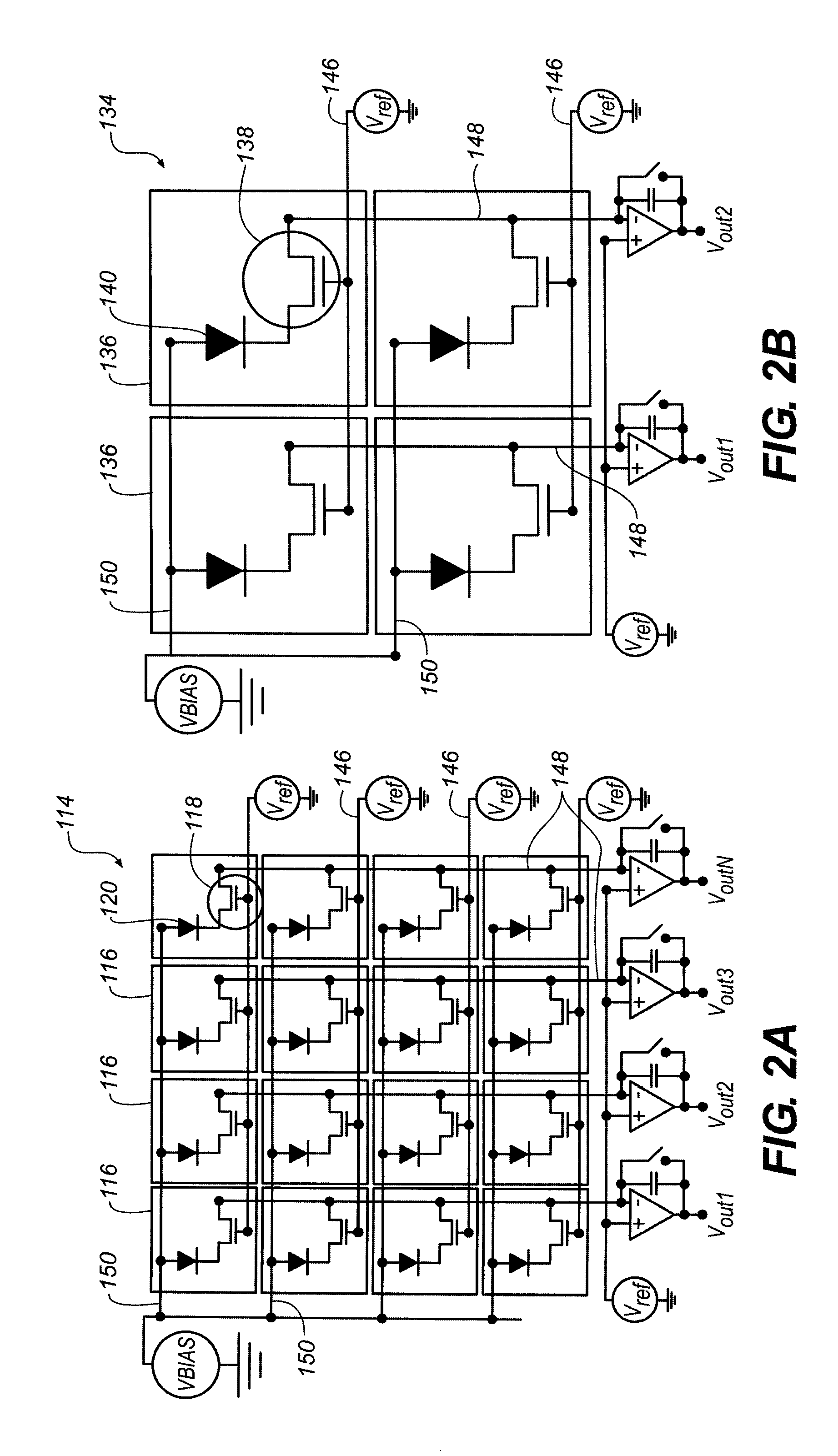
[0031]The first detector 110 includes a first substrate 112 upon which a first detector array 114 is disposed. The first detector array 114 preferably includes a number of first pixels 116, and each first pixel 116 includes a first readout element 118 and a first photosensitive element 120. A first scintillating phosphor screen 122 is disposed on the first detector array 114, on a side opposite the first substrate 112. A ...


PUM


Abstract

Embodiments of radiographic imaging apparatus and methods for operating the same can include a first scintillator, a second scintillator, a plurality of first photosensitive elements, and a plurality of second photosensitive elements. The plurality of first photosensitive elements receives light from the first scintillator and has first photosensitive element characteristics chosen to cooperate with the first scintillator properties. The plurality of second photosensitive elements are arranged to receive light from the second scintillator and has second photosensitive element characteristics different from the first photosensitive element characteristics and chosen to cooperate with the second scintillator properties. Further, the first scintillator can have first scintillator properties and the second scintillator can have second scintillator properties different from the first scintillator properties.

Description

BACKGROUND OF THE INVENTION[0001]1. Technical Field[0002]The present invention relates to digital radiographic detectors. More particularly, the present invention relates to a digital radiographic detector with two scintillating screens and improved spatial sampling.[0003]2. Description of Related Art[0004]Digital radiographic imaging devices are becoming increasingly used in both medicinal and industrial applications. Conventional radiographic imaging devices generally include an array of pixels formed on a substrate. Each pixel includes a photosensitive element and a readout element. Conventionally known photosensitive elements include p-n junction photodiodes, metal-insulator-semiconductor (MIS) photo-capacitors, and pinned photodiodes, among others. The presently most commonly used readout element is a thin-film transistor (TFT), although other readout elements also may be used. In use, the photosensitive element converts an incident light into an electrical signal, and the elec...

Claims


Application Information

Patent Timeline
IPC IPC(8): G01T1/20
CPCG01T1/2018G01T1/20184G01T1/20181
Inventor TREDWELL, TIMOTHY J.SHAFER, MARK E.KULPINSKI, ROBERT W.WOJCIK, TIMOTHY J.
Owner CARESTREAM HEALTH INC
Why Eureka
Try Eureka