
Diagnosis of benign and cancerous growths by measuring circulation tumor stem cells and serum annexina2

a technology of stem cells and tumors, applied in the field of diagnosis, can solve the problems of poor efficiency of such tests, no diagnostic tests have been described to predict the presence of pre-cancerous, benign tumors (such as colon polyps) in patients, and achieve the effect of increasing the tumorigenic and metastatic potential of cells and larger tumors

Active Publication Date: 2012-02-09
BOARD OF RGT UNIV OF TEXAS SYST THE
View PDF1 Cites 10 Cited by
  • Summary
  • Abstract
  • Description
  • Claims
  • Application Information

AI Technical Summary

Benefits of technology

[0023]FIGS. 8A-8D show that PG over-expression in HEK-mGAS cells increases tumorigenic and metastatic potential of the cells in vivo. FIG. 8A: HEK-C and HEK-mGAS cells were subdermally inoculated into the flank of the athymic nude mice. As can be seen, HEK-mGAS grew significantly larger tumors compared to the HEK-C control cells. FIG. 8B: Table is presented. Only 1 out 6 mice inoculated with HEK-C cells grew tumors whereas all 6 out 6 mice inoculated with HEK-mGAS cells grew as tumors. HCT-116, which is a human colorectal cancer cell line, was used as a positive control. FIG. 8C. HEK-mGAS cells were inoculated into the spleen of the nud...

Problems solved by technology

Developing relatively simple, non-invasive methods for diagnosing the presence of either benign tumors, such as colonic hyperplasia, adenomatous polyps, or cancerous tumors, such as adenocarcinomas at different stages of the disease, has remained a challenge.
Efficiency of such tests are poor, because of the rare presence of cancer cells within a very large number of blood cells (1-1000 CTCs in 109 blood cells / ml blood).
The presence of CTCs in the blood will not detect the presence of benign tumors at a pre-cancerous stage, such as adenomatous polyps growing in the colon of the patients.
So while one can potentially diagnose and predict the presence of cancerous tumors using the newly described assays for either circulating tumor cells or cancer specific antigens, such as Her / neu, PSA or AnnexinA2, no diagnostic tests have been described to predict the presence of pre-cancerous, benign tumors (such as colon polyps) in the patients.

Method used


Image

  • Diagnosis of benign and cancerous growths by measuring circulation tumor stem cells and serum annexina2
  • Diagnosis of benign and cancerous growths by measuring circulation tumor stem cells and serum annexina2
  • Diagnosis of benign and cancerous growths by measuring circulation tumor stem cells and serum annexina2

Examples


example 1

Autocrine and Endocrine Growth Factors Mediate Tumorigenic Effects on Epithelial Cells by Binding Cell-Surface Associated AnnexinA2 (CS-ANXA2)

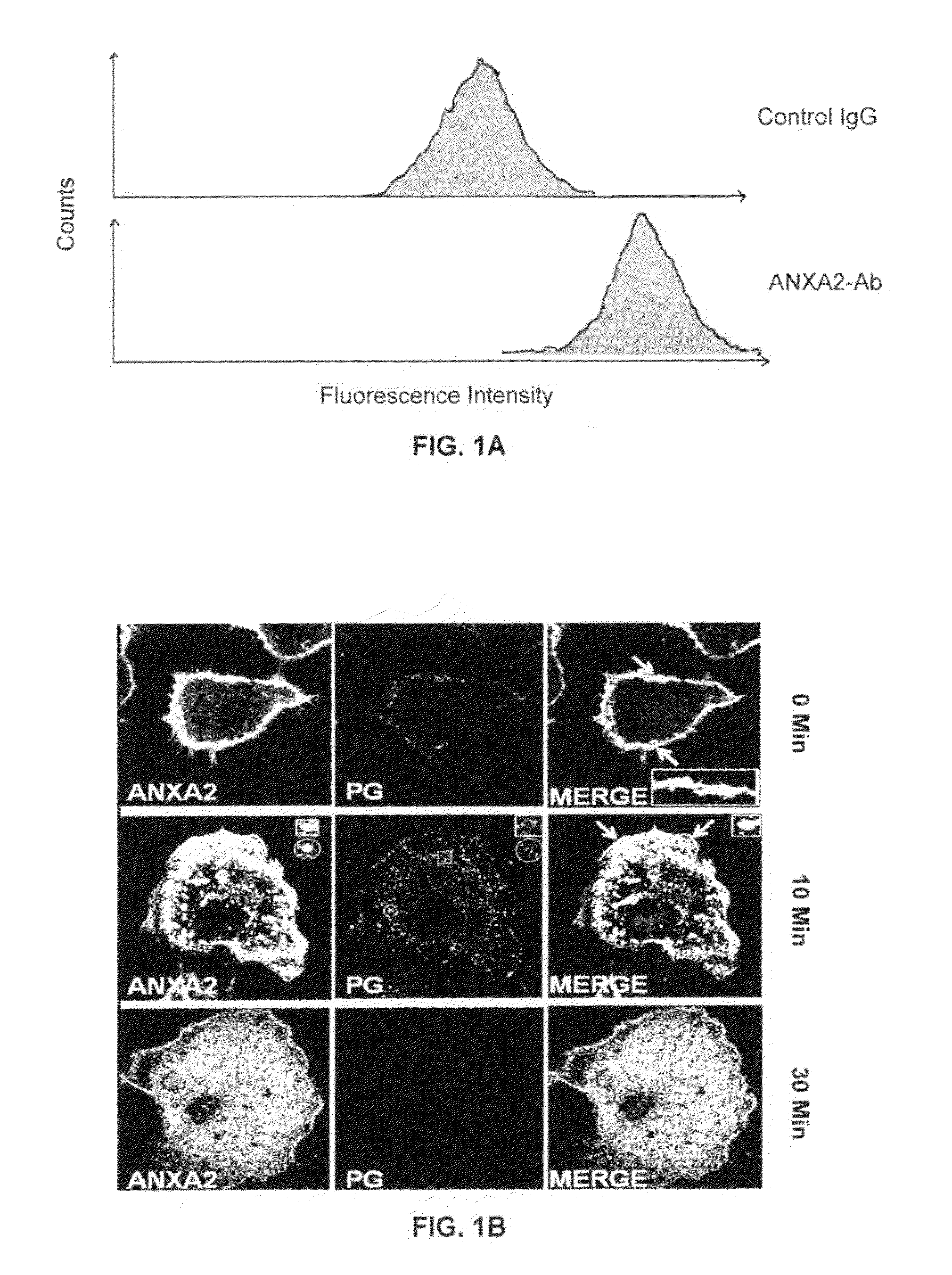
[0053]Non-amidated precursor forms of gastrins, called progastrin peptides (PG), are potent mitogens, and exert proliferative / anti-apoptotic / co-carcinogenic and tumorigenic effects on normal and cancerous intestinal, pancreatic and lung epithelial cells (22-29). Cell surface associated ANXA2 (CS-ANXA2) functions as a high affinity, non-conventional receptor protein for PG peptides (30), and is required for mediating the growth promoting effects of PG on epithelial cells from the intestine, embryonic kidney and pancreatic cancers, in vitro (27,30) and in vivo (31). Data demonstrating the presence of CS-ANXA2 on intestinal epithelial cells is presented in FIGS. 1A-1B. The fluorescence intensity of cells labeled with anti-ANXA2-antibodies (Abs) was increased >10-fold compared to cells labeled with control IgG (FIG. 1A), confirming the presence of...

example 2

AnnexinA2 Expression is Required for Measuring an Increase in Stem Cell Populations

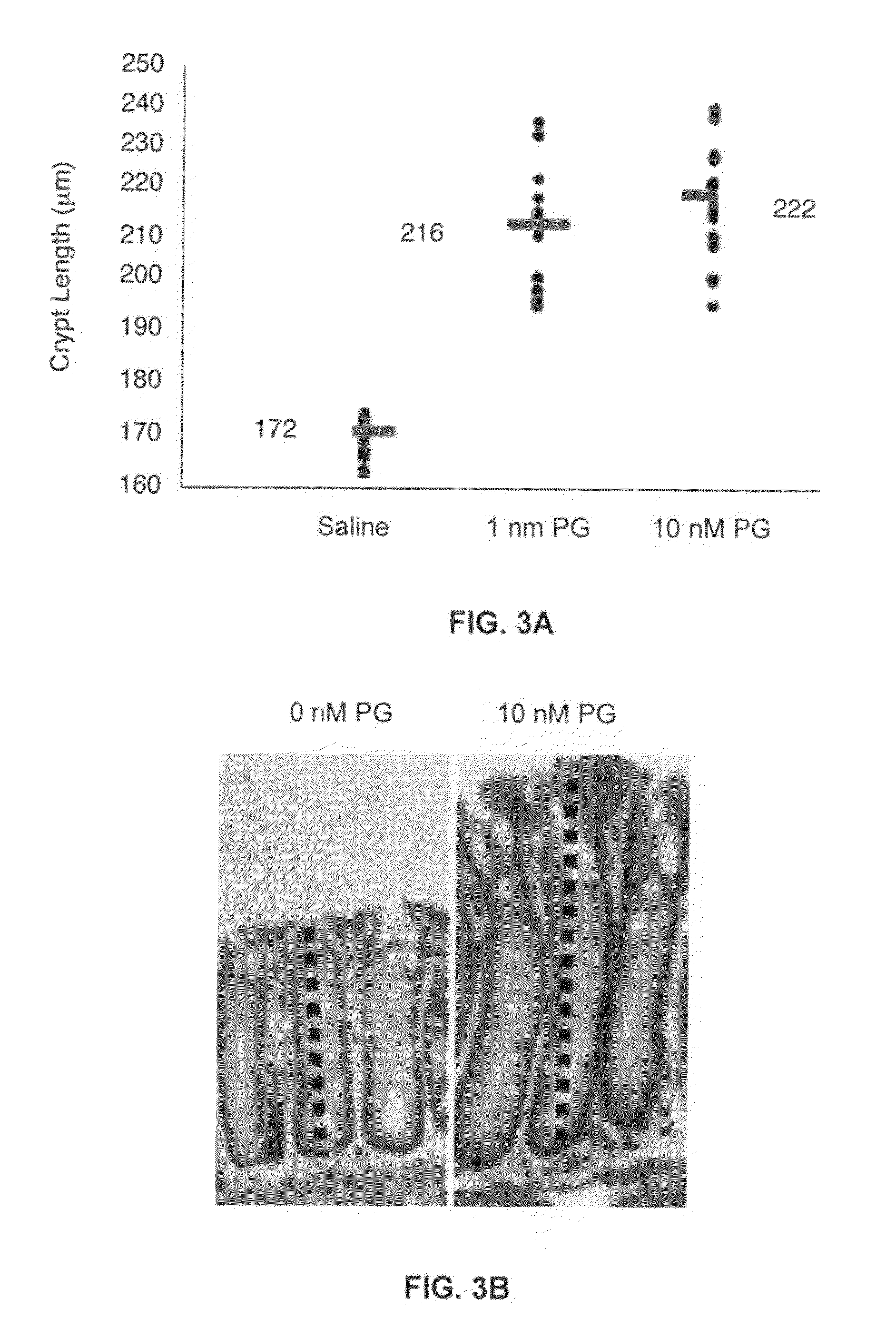
[0054]The proliferative and tumorigenic effects of PG peptides are associated with the significant increase in the stem cell populations within colonic crypts of mice (31). A significant increase in the length of the colonic crypts in response to 1-10 nM PG was confirmed in C57Bl / J6 mice (FIGS. 3A-3B), as reported with transgenic FVB / N mice (25). The concentrations of stem cell markers, CD44 and DCAMKL−1 were significantly increased in colonic crypts of mice treated with 10 nM PG (FIG. 3C), and the total number of cells positive for CD44 and DCAMKL+1 markers were also significantly increased in the colonic crypts of PG-treated mice (FIG. 3D). ANXA2 knockout (ANXA2− / −) mice were used to examine if ANXA2 expression was required for mediating the proliferative effects of PG on the colonic crypts of mice. The ANXA2− / − mice were confirmed by Western Blot analysis (FIG. 4A). Unlike the wild type ANXA2+ / + mi...

example 3

[0056]Tumorigenic Transformation of Kidney Epithelial Cells by Over-Expression of PG, is Associated with Significant Up-Regulation of ANXA2 and Stem Cell Marker Proteins

[0057]In order to further examine the tumorigenic and co-carcinogenic effects of PG / ANXA2, non-transformed embryonic kidney epithelial cells (HEK-293) were used since these cells are very easy to clone and are responsive to growth effects of PG peptides. Clones of HEK-293 cells were generated which either expressed the control vector (HEK-C) or the human gastrin gene vector (HEK-mGAS). The clones over-expressing gastrin gene were confirmed to be over-expressing the PG peptide (9 Kd) (FIG. 6A). Over-expression of PG in HEK-293 cells induced activation of NFκB and β-catenin pathways (including p65Ser276, COX-II, β-catenin, c-Myc, cyclin D1) (FIGS. 6B-6E) (31). Over-expression of PG in HEK-293 cells also up-regulated the expression of stem cell markers DCAMKL−1, CD44 and Lgr5 in the HEK-mGAS vs the HEK-C cells as shown ...


PUM


Abstract

Provided herein is a method for diagnosing / prognosing a metastatic cancer in a subject by measuring and detecting one or more of CS-ANXA2, DCAMKL, Lgr5 or CS-ANAX2 and DCAMKL or CS-ANXA2 and Lgr5 positive circulating tumor stem cells in the subject's blood or plasma. Also provided is a method for distinguishing the presence of early stage primary cancer from advanced stage metastatic cancer in the subject by measuring and detecting AnnexinA2, CS-ANXA2 and DCAMKL−1 or Lgr5 in the blood or plasma. In addition, there is provided a method for distinguishing the presence of benign, pre-cancerous tumorous growths or cancerous tumors in the subject by measuring and detecting AnnexinA2 and circulating tumor stem cells positive for CS-ANXA2 and DCAMKL or CS-ANXA2 and Lgr5 in the blood or plasma.

Description

CROSS-REFERENCES TO RELATED APPLICATION[0001]This nonprovisional application claims benefit of priority under 35 U.S.C. §119(e) of provisional U.S. Ser. No. 61 / 398,642, filed Jun. 28, 2010, now abandoned, and U.S. Ser. No. 61 / 398,644, filed Jun. 28, 2010, now abandoned, the entirety of both of which are hereby incorporated by reference.FEDERAL FUNDING[0002]This invention was made with government support under Grant Number R01CA097959 awarded by the National Institutes of Health. The government has certain rights in the invention.BACKGROUND OF THE INVENTION[0003]1. Field of the Invention[0004]The present invention relates generally to the fields of detection and diagnosis and, more specifically, to diagnosis of the presence of early stage (primary) or late stage (metastatic) cancerous tumors, using a combinatorial approach of measuring circulating tumor stem cells and serum AnnexinA2.[0005]2. Description of the Related Art[0006]Developing relatively simple, non-invasive methods for d...

Claims


Application Information

Patent Timeline
IPC IPC(8): G01N33/574
CPCG01N2333/4718G01N33/57492
Inventor SINGH, POMILA
Owner BOARD OF RGT UNIV OF TEXAS SYST THE
Learn More