Method and System for Image Based Device Tracking for Co-registration of Angiography and Intravascular Ultrasound Images

a technology of intravascular ultrasound and image based device, which is applied in the field of image based device detection and tracking for co-registration of angiographic fluoroscopic images and intravascular ultrasound images, can solve problems such as difficulty in fully understanding the spatial structure of the vessel

Active Publication Date: 2012-03-08
SIEMENS HEALTHCARE GMBH
View PDF1 Cites 144 Cited by
  • Summary
  • Abstract
  • Description
  • Claims
  • Application Information

AI Technical Summary

Problems solved by technology

However, the lack of vessel orientation information in IVUS images m...

Method used


Image

  • Method and System for Image Based Device Tracking for Co-registration of Angiography and Intravascular Ultrasound Images
  • Method and System for Image Based Device Tracking for Co-registration of Angiography and Intravascular Ultrasound Images
  • Method and System for Image Based Device Tracking for Co-registration of Angiography and Intravascular Ultrasound Images

Examples


Embodiment Construction

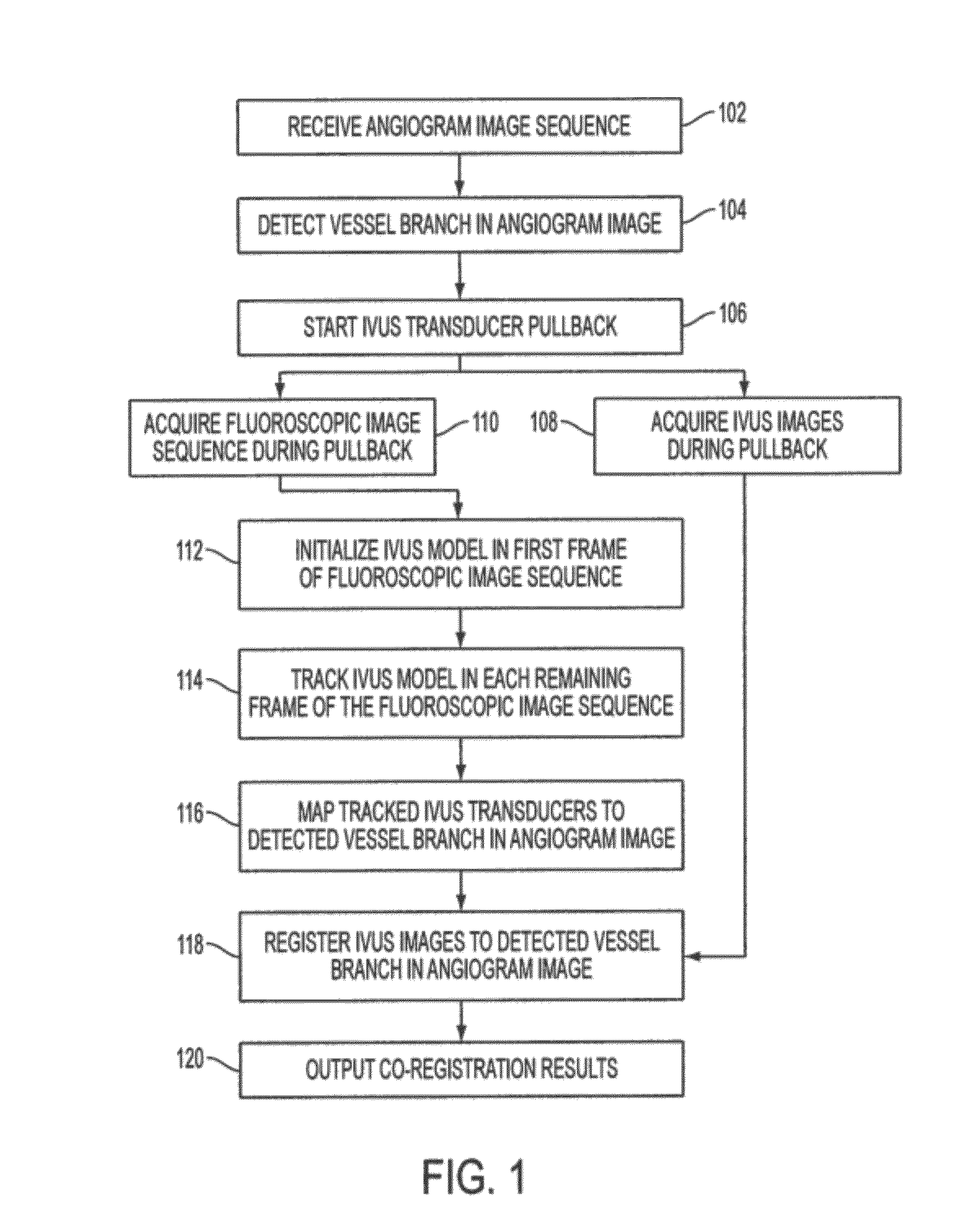
[0020]The present invention relates to a method and system for co-registration of angiography and Intravascular Ultrasound (IVUS) images using image based device tracking. Embodiments of the present invention are described herein to give a visual understanding of the co-registration method. A digital image is often composed of digital representations of one or more objects (or shapes). The digital representation of an object is often described herein in terms of identifying and manipulating the object. Such manipulations are virtual manipulations accomplished in the memory or other circuitry / hardware of a computer system. Accordingly, is to be understood that embodiments of the present invention may be performed within a computer system using data stored within the computer system.

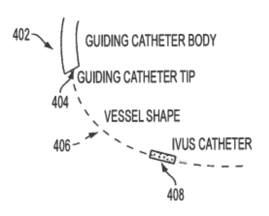
[0021]Registration of IVUS and angiography images finds the position of each IVUS image plane along a vessel branch during the IVUS pullback. Manuel labeling, which has previously been used to specify the ...


PUM


Abstract

A method and system for co-registration of angiography data and intra vascular ultrasound (IVUS) data is disclosed. A vessel branch is detected in an angiogram image. A sequence of IVUS images is received from an IVUS transducer while the IVUS transducer is being pulled back through the vessel branch. A fluoroscopic image sequence is received while the IVUS transducer is being pulled back through the vessel branch. The IVUS transducer and a guiding catheter tip are detected in each frame of the fluoroscopic image sequence. The IVUS transducer detected in each frame of the fluoroscopic image sequence is mapped to a respective location in the detected vessel branch of the angiogram image. Each of the IVUS images is registered to a respective location in the detected vessel branch of the angiogram image based on the mapped location of the IVUS transducer detected in a corresponding frame of the fluoroscopic image sequence.

Description

[0001]This application claims the benefit of U.S. Provisional Application No. 61 / 359,431, filed Jun. 29, 2010, the disclosure of which is herein incorporated by reference.BACKGROUND OF THE INVENTION[0002]The present invention relates to image based device detection and tracking, and more particularly, image based device detection and tracking for co-registration of angiographic fluoroscopic images and intravascular ultrasound images.[0003]Angiography and Intravascular ultrasound (IVUS) are two widely used modalities to image coronary vessels in coronary interventions. Both modalities have various advantages. X-ray angiography is a modality that is commonly used to guide percutaneous coronary interventions (PCI). IVUS is an imaging technique in which an ultrasound transducer is attached to an end of a guidewire. The guidewire is first guided through a guiding catheter and from there through the vessels of a patient. The ultrasound transducer is used to obtain ultrasound images from w...

Claims


Application Information

Patent Timeline
IPC IPC(8): A61B6/00A61B8/00
CPCA61B6/00A61B6/12A61B6/481A61B6/487A61B6/504G06T2207/10132A61B8/0891A61B8/12A61B8/5238G06T7/2046G06T2207/10121A61B6/5247G06T7/251
Inventor WANG, PENGPRUMMER, SIMONECHEN, TERRENCECOMANICIU, DORINECABERT, OLIVIEROSTERMEIER, MARTIN
Owner SIEMENS HEALTHCARE GMBH
Why Eureka
Try Eureka