
Transdermal Sampling and Analysis Device

a technology which is applied in the field of transdermal sampling and analysis device, can solve the problems of inconvenient procedure, inconvenient operation, and inability to accurately determine the effect of the procedure, and achieve the effect of altering the permeability of the stratum corneum

Active Publication Date: 2012-06-14
CLINITECH LLC +2
View PDF13 Cites 30 Cited by
  • Summary
  • Abstract
  • Description
  • Claims
  • Application Information

AI Technical Summary

Benefits of technology

"The patent describes a method and apparatus for safely and non-invasively extracting biological samples through the skin using a disruptor unit that generates heat to alter the permeability of the stratum corneum without damaging it. This allows interstitial fluid to flow and be collected for analysis. The method and apparatus can be used in various configurations and with different materials, and may also include a disposable applicator unit for easy replacement. The sensed property values of the biological sample can be displayed to the user or communicated to a remote computer / server for analysis and monitoring. The technical effects of the patent include a safer and non-invasive method for extracting biological samples and the potential for remote analysis and monitoring."

Problems solved by technology

Conventional methods for obtaining biological samples are typically painful and invasive.
This procedure may be uncomfortable, painful, and especially irritating when it has to be performed multiple times a day in the case of diabetics.
In addition to pain and discomfort, there are other undesirable side effects associated with these invasive tissue extraction techniques.
For example, diabetics who also suffer from hemophilia face the danger of severe hemorrhage every time they have to test their blood glucose levels using invasive procedures.
In another example, these invasive procedures expose immuno-compromised diabetics to increased chances of local or systemic infections.
To obtain these larger biological samples, painful and invasive procedures must be employed, which are not desirable.

Method used


Image

  • Transdermal Sampling and Analysis Device
  • Transdermal Sampling and Analysis Device
  • Transdermal Sampling and Analysis Device

Examples


Embodiment Construction

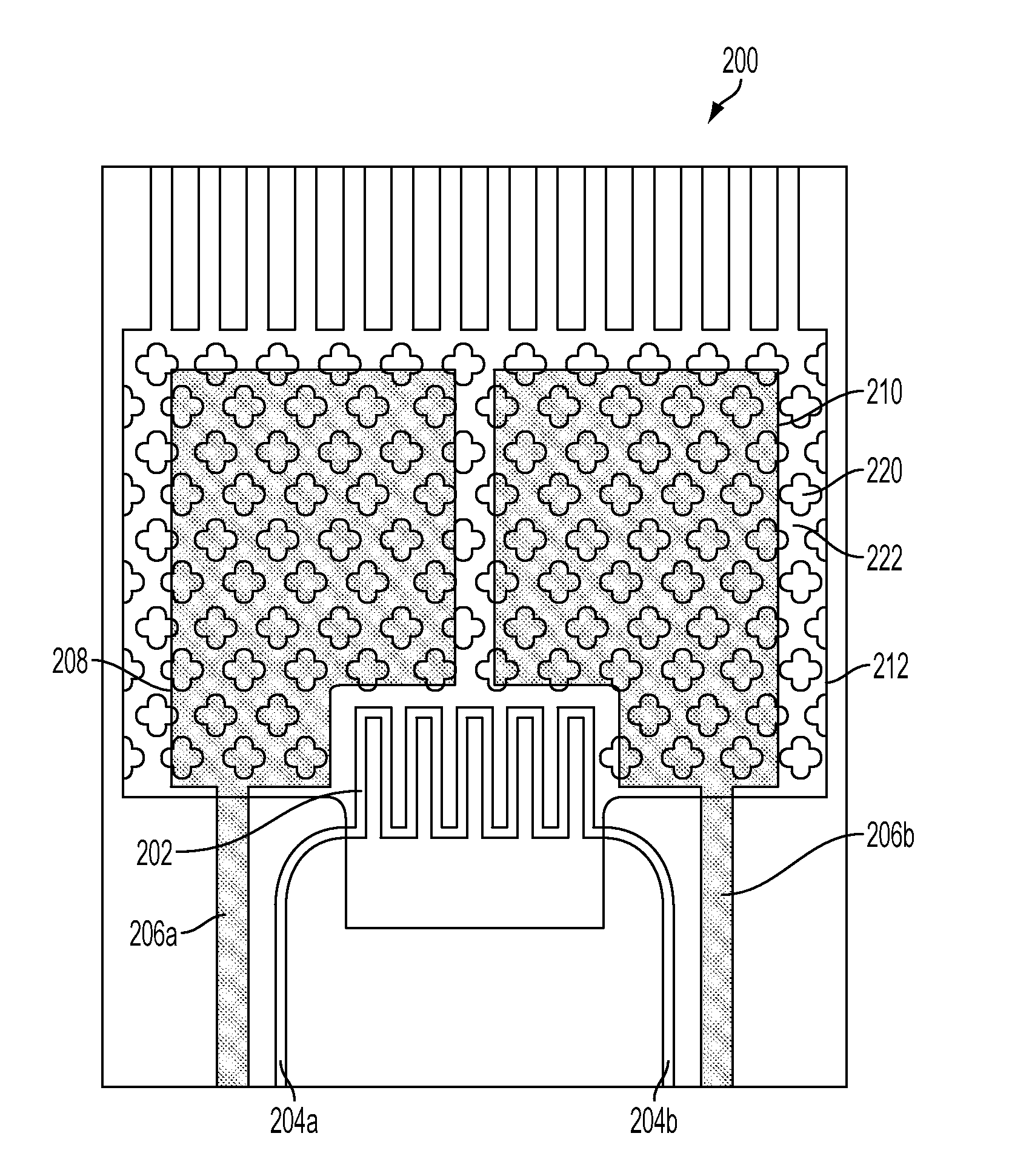
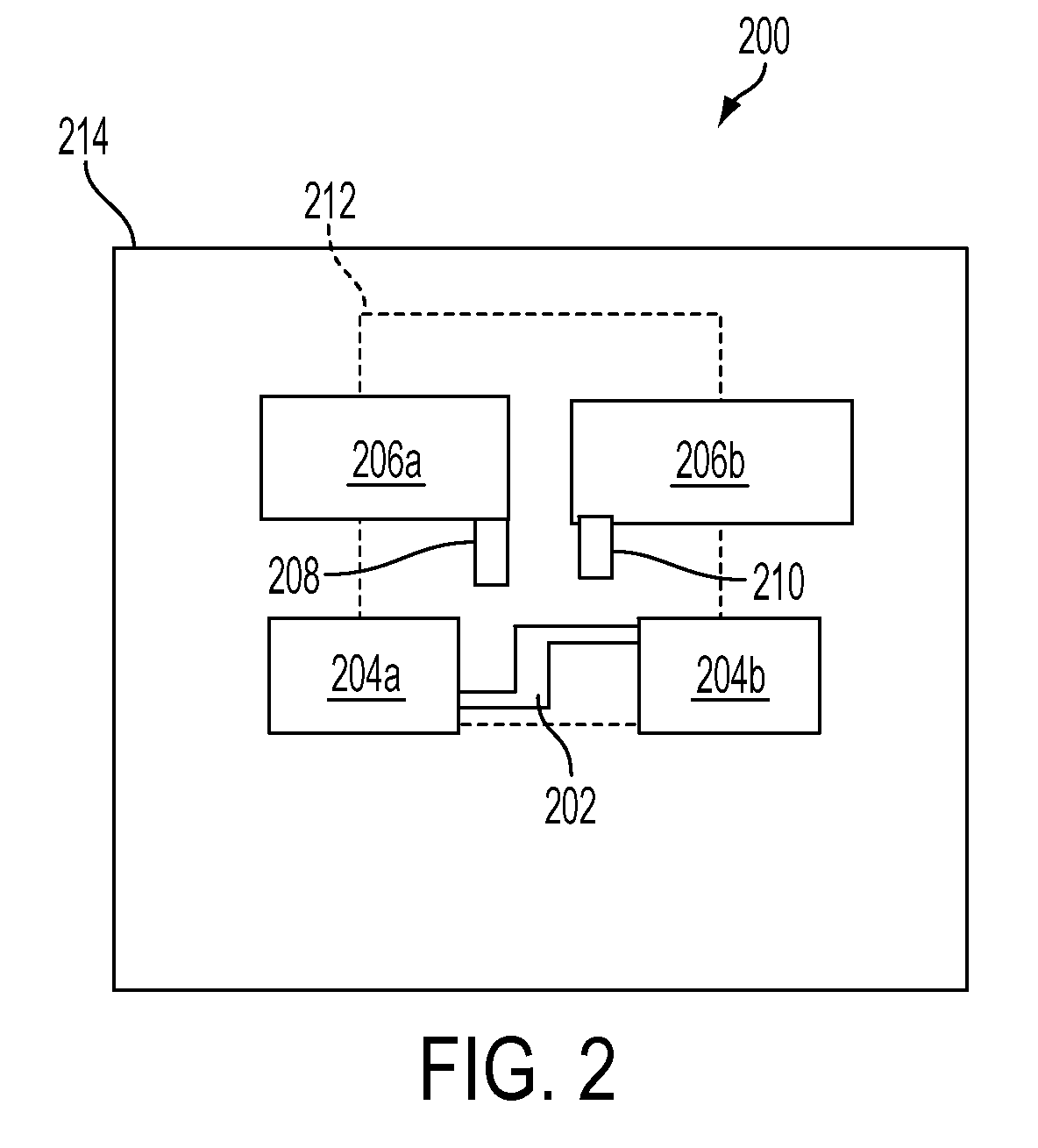
[0038]The various embodiments will be described in detail with reference to the accompanying drawings. Wherever possible, the same reference numbers will be used throughout the drawings to refer to the same or like parts. References made to particular examples and implementations are for illustrative purposes, and are not intended to limit the scope of the invention or the claims.

[0039]Conventional methods for obtaining biological samples from a subject are invasive, uncomfortable, and painful. For example, conventional glucose biosensors require that diabetics obtain blood samples by puncturing or lacerating their skin using a sharp blade or pin. The blood sample may then collected and delivered to a biosensor which detects glucose levels of the sample blood. While such biosensors are often marketed as being “pain-free,” users often experience some degree of discomfort that they may become inured to over repeated samplings. Regardless, conventional biosensors may cause discomfort, ...


PUM


Abstract

Transdermal sampling and analysis device, method and system are provided for non-invasively and transdermally obtaining biological samples from a subject and determining levels of analytes of the obtained biological samples. The transdermal sampling and analysis device, method and system may cause disruption to the skin cells to create capillary-like channels from which biological samples may flow to the transdermal sampling and analysis device. The transdermal sampling and analysis device, method and system may collect the biological samples in a reservoir where the biological sample may chemically react with a biologically reactive element. A sensor may convert the produced electrons (ions) into measured electrical signals. The converted signals may be measured and the levels of an analyte may be determined based on the measured signals.

Description

RELATED APPLICATIONS[0001]This application claims the benefit of priority to U.S. Provisional Patent Application Ser. No. 61 / 421,982 entitled “Transdermal Sampling and Analysis Device,” filed on Dec. 10, 2010, the entire contents of which are hereby incorporated by reference.BACKGROUND[0002]Effective diagnoses and treatment of diseases depend on accurate monitoring of the current physiological state of a subject. Monitoring the concentration of molecules in the body is an example of a method for determining the physiological state. For example, diabetics must actively monitor their body's glucose levels to treat and prevent potentially life threatening conditions such as hypo- or hyperglycemia.[0003]Monitoring the internal physiological state of a subject requires a two step process. First, biological samples must be obtained from the body of the subject. Second, the sample must be analyzed using any of a variety of methods and systems. Some common methods and systems that may be us...

Claims


Application Information

Patent Timeline
Patent Type & Authority Applications(United States)
IPC IPC(8): A61B5/157
CPCA61B5/0022A61B2562/0295A61B5/1486A61B5/150022A61B5/150076A61B5/150122A61B5/150358A61B5/150412A61B5/150854A61B5/150862A61B5/15087A61B5/150946A61B5/150984A61B5/15134A61B5/157A61B5/14532
Inventor CURRIE, JOHN F.MARCANIO, JOSEPH A.VIDALIS, JOSEPH J.NADARAJAN, SUNDAR B.
Owner CLINITECH LLC
Learn More