Detection of Landmarks and Key-frames in Cardiac Perfusion MRI Using a Joint Spatial-Temporal Context Model

a spatial-temporal context and landmark detection technology, applied in image analysis, image enhancement, instruments, etc., can solve the time-consuming problems of clinical routine applications of cardiac perfusion mri, and achieve the effect of enhancing the discriminative capabilities of the contextual model

Inactive Publication Date: 2012-07-12
SIEMENS HEALTHCARE GMBH +1
View PDF17 Cites 17 Cited by
  • Summary
  • Abstract
  • Description
  • Claims
  • Application Information

AI Technical Summary

Benefits of technology

[0009]In an exemplary embodiment of the present invention, instead of utilizing spatial context only or both spatial and temporal contexts separately, there is provided a joint spatial-temporal context model (also referred to as “the contextual model”) under a learning-based object detection framework to automatically detect anatomic landmarks and identify key-frames (a key-frame could be different for different heart chambers) simultaneously. The contextual model is applied to a three-dimensional (3D) volume reconstructed from an original perfusion magnetic resonance imaging (MRI) sequence. In addition, because each individual landmark has limited local evidence to identify, context is constructed not only based on individual landmarks (also referred to as “the target” or “the target landmarks”), but also landmarks that are correlated to the target. Taking into account correlations among landmarks and across the temporal dimension includes more evidence and enhances the discriminative capabilities of the contextual model. The target landmarks may include right ventricle (RV) insertion points and left ventricle (LV) center. And the key-frames may be identified for both RV and LV.

Problems solved by technology

However, clinical routine applications of cardiac perfusion MRI are time-consuming due to the analysis of perfusion data.

Method used


Image

  • Detection of Landmarks and Key-frames in Cardiac Perfusion MRI Using a Joint Spatial-Temporal Context Model
  • Detection of Landmarks and Key-frames in Cardiac Perfusion MRI Using a Joint Spatial-Temporal Context Model
  • Detection of Landmarks and Key-frames in Cardiac Perfusion MRI Using a Joint Spatial-Temporal Context Model

Examples


Embodiment Construction

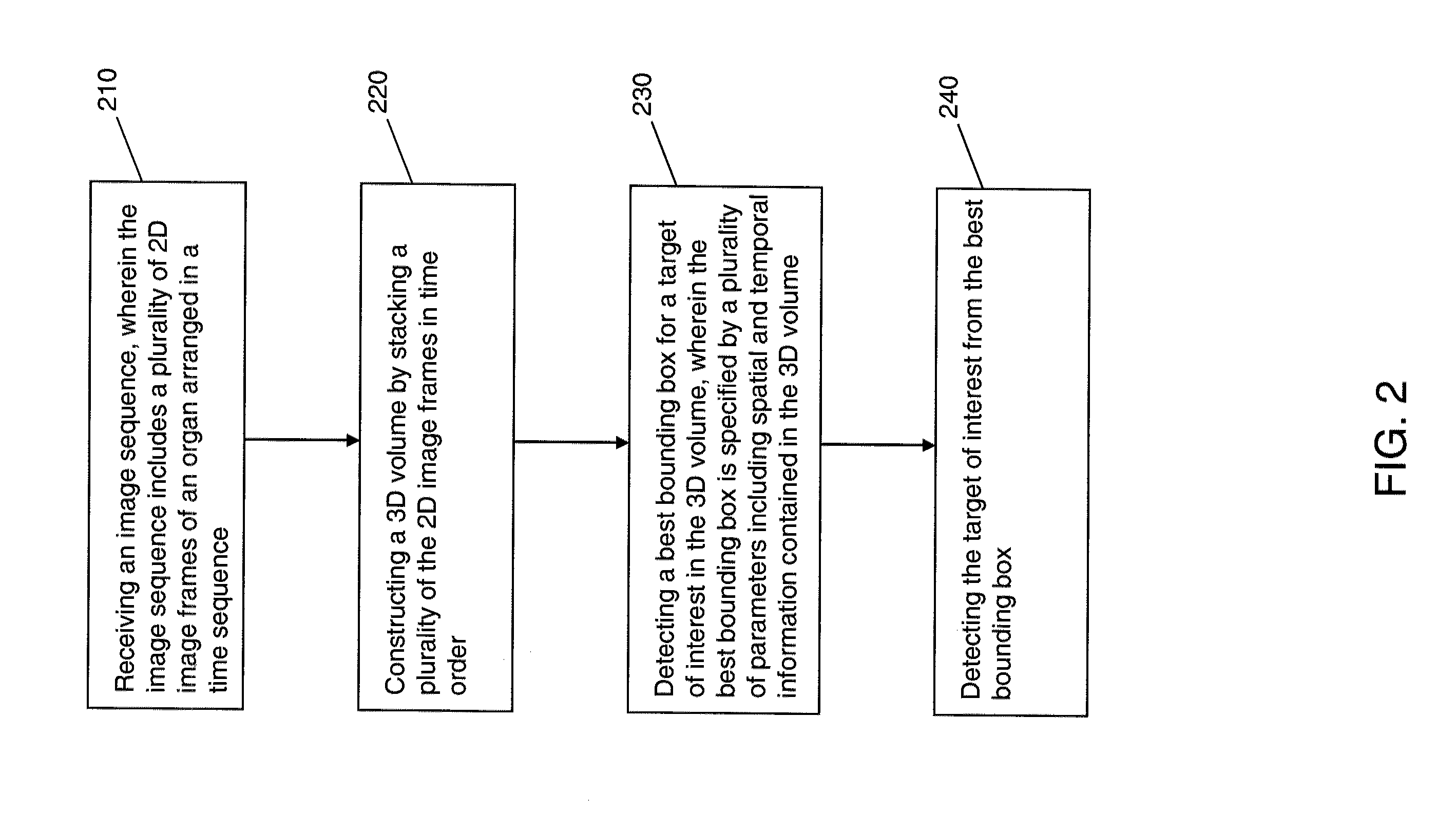
[0032]An overall workflow of a method for the detection of landmarks and key-frames in a cardiac perfusion magnetic resonance imaging (MRI) sequence using a joint spatial-temporal context model, according to an exemplary embodiment of the present invention, will now be discussed, in brief, with primary reference to FIG. 2. Detailed explanations of the workflow in FIG. 2 will then follow with reference to the remainder of the figures.

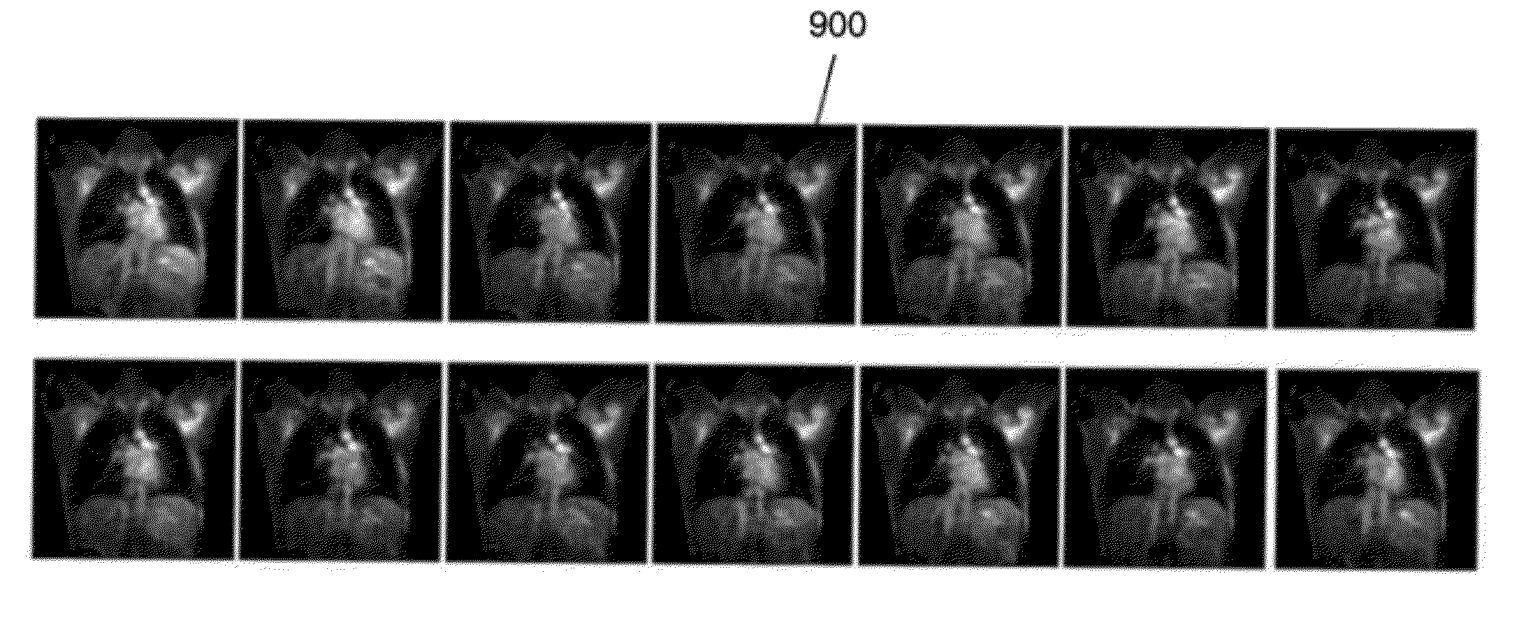
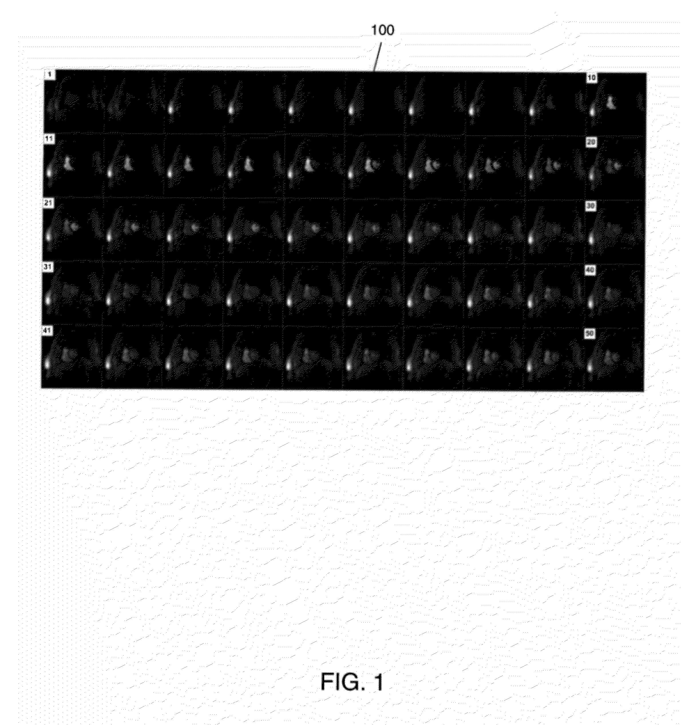
[0033]The following discussion formulates the simultaneous landmark and key-frame detection of FIG. 2 into a three-dimensional (3D) object detection framework. Referring to FIG. 2, an image sequence is received (210). The image sequence includes a plurality of two-dimensional (2D) image frames of an organ arranged in a time sequence. An example of the received image sequence may be the cardiac perfusion MRI sequence 100 of FIG. 1. However, the present invention is not limited thereto. For example, the received image sequence may be chest MRI sequence 900...


PUM


Abstract

A method including receiving an image sequence, wherein the image sequence includes a plurality of two-dimensional (2D) image frames of an organ arranged in a time sequence; constructing a three-dimensional (3D) volume by stacking a plurality of the 2D image frames in time order; detecting a best bounding box for a target of interest in the 3D volume, wherein the best bounding box is specified by a plurality of parameters including spatial and temporal information contained in the 3D volume; and determining the target of interest from the best bounding box.

Description

CROSS-REFERENCE TO RELATED APPLICATION[0001]This application claims priority under 35 U.S.C. §119 to U.S. Provisional Application No. 61 / 385,180, filed Sep. 22, 2010, the disclosure of which is incorporated by reference herein in its entirety.BACKGROUND OF THE INVENTION[0002]1. Technical Field[0003]The present invention relates to cardiac perfusion magnetic resonance imaging (MRI), and more particularly, to the automated detection of anatomic landmarks and key-frames from perfusion MRI sequences.[0004]2. Discussion of the Related Art[0005]Myocardial first-pass perfusion cardiovascular MRI has significantly advanced over the last decade and has shown a great deal of value in clinical applications for diagnosis and prognosis in heart diseases. However, clinical routine applications of cardiac perfusion MRI are time-consuming due to the analysis of perfusion data.[0006]Precise information on both morphology and functions of the left ventricle (LV) and the right ventricle (RV) is helpfu...

Claims


Application Information

Patent Timeline
Patent Type & Authority Applications(United States)
IPC IPC(8): G06K9/00
CPCG06K9/4609G06K2209/051G06T7/0081G06T2207/30048G06T2207/10088G06T2207/20144G06T2207/20164G06T7/0087G06T7/11G06T7/143G06T7/194G06V10/443G06V2201/031
Inventor LU, XIAOGUANGGUHRING, JENSXUE, HUIJOLLY, MARIE-PIERREGUETTER, CHRISTOPHGEORGESCU, BOGDANZUEHLSDORFF, SVENLITTMANN, ARNE
Owner SIEMENS HEALTHCARE GMBH
Why Eureka
Try Eureka