Methods and apparatus for diagnosis and/or prognosis of cancer

a cancer and prognosis technology, applied in the field of detection, diagnosis and/or prognosis of cancers, can solve the problems of high variability in the prognosis of stage i lung cancer, and clinical techniques that cannot distinguish stage i patients into long-term survivors and short-term survival groups, so as to achieve detection, diagnosis and/or prognosis.

Active Publication Date: 2012-07-12
H LEE MOFFITT CANCER CENT & RES INST INC
View PDF3 Cites 34 Cited by
  • Summary
  • Abstract
  • Description
  • Claims
  • Application Information

AI Technical Summary

Benefits of technology

[0005]The subject invention concerns methods and apparatus for the detection, diagnosis, and/or prognosis of cancer in a person or animal by analyzing centrosomal features. In one embodiment, a method of the invention includes receiving an image of one or more cells; selecting a region of interest in one cell; segmenting the region of interest in the one cell to delineate or isolate at least one centrosome; extracting one or more features from the segmented image; and analyzing the extracted features to diagnose cancer. In another embodiment, the progression of cancer can be predicted through analysis of the extracted features. In one embodiment, methods of the invention can be performed by a quantitative cancer analysis system including a diagnosis module and/or a prognosis module. In one embodiment, the method can be performed using an image processing system. Methods of the invention can be used to distinguish longer term can

Problems solved by technology

Unfortunately, the prognosis of stage I lung cancer is highly variable.
In our study of patients with stage I lung cancer, cl

Method used


Image

  • Methods and apparatus for diagnosis and/or prognosis of cancer
  • Methods and apparatus for diagnosis and/or prognosis of cancer
  • Methods and apparatus for diagnosis and/or prognosis of cancer

Examples


example 1

Quantificational and Statistical Analysis of the Differences in Centrosomal Features of Untreated Lung Cancer Cells and Normal Cells

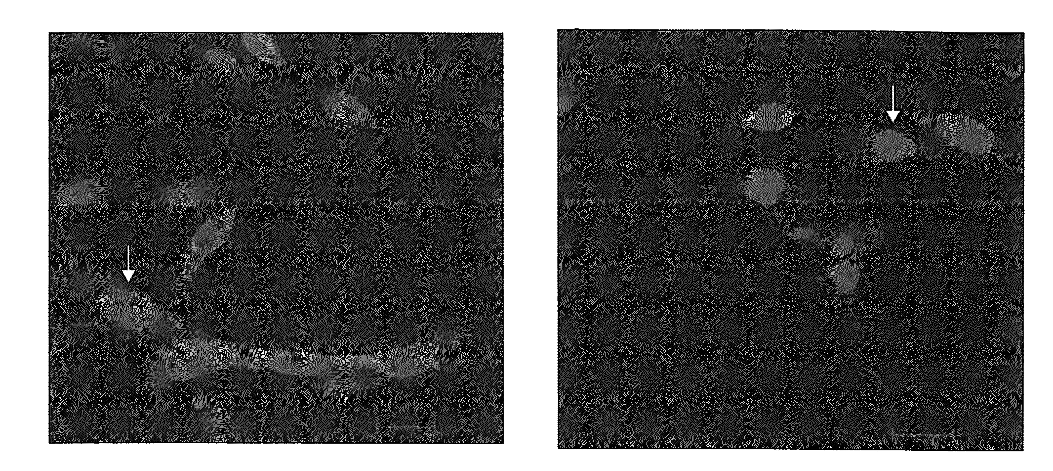
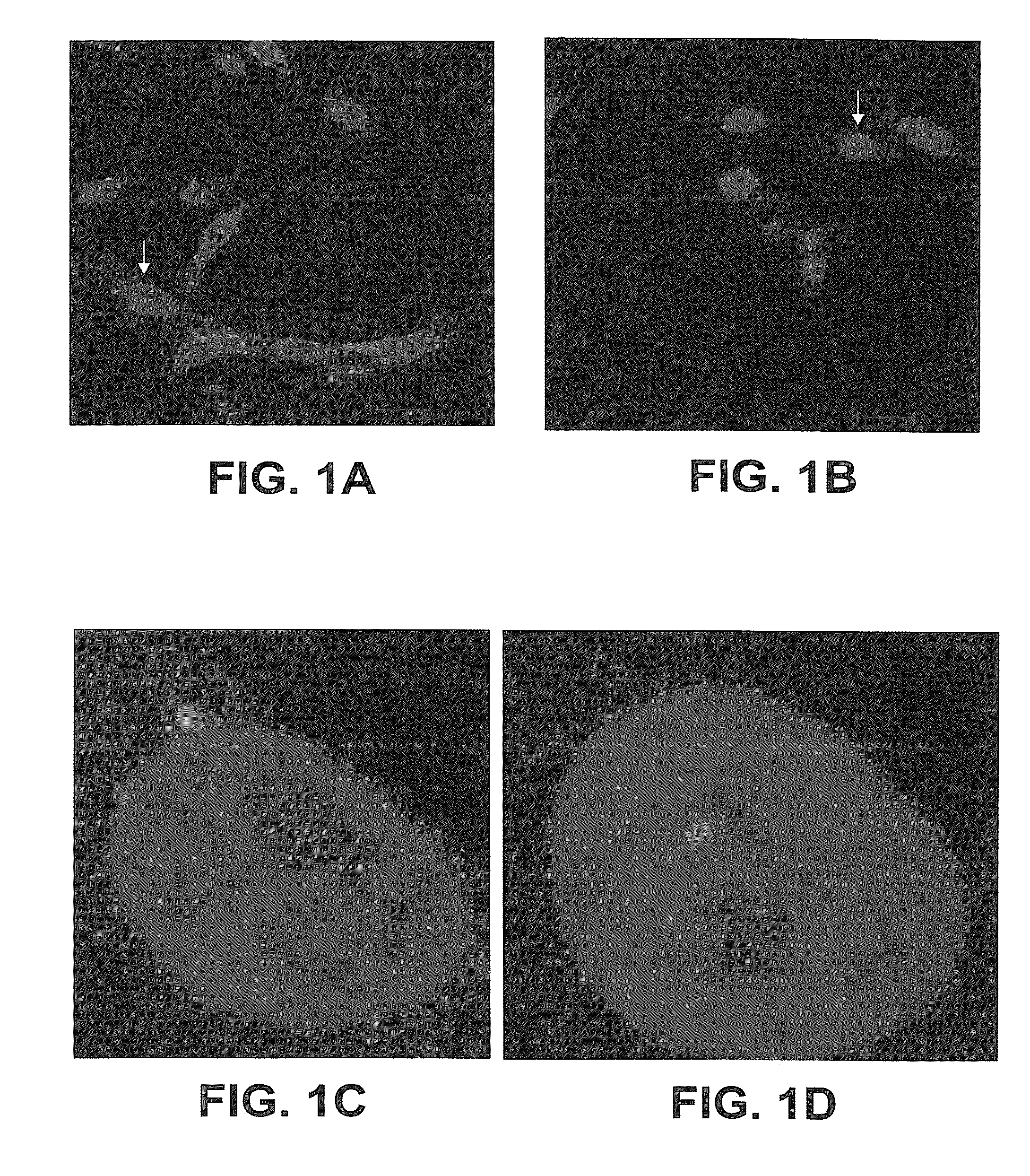
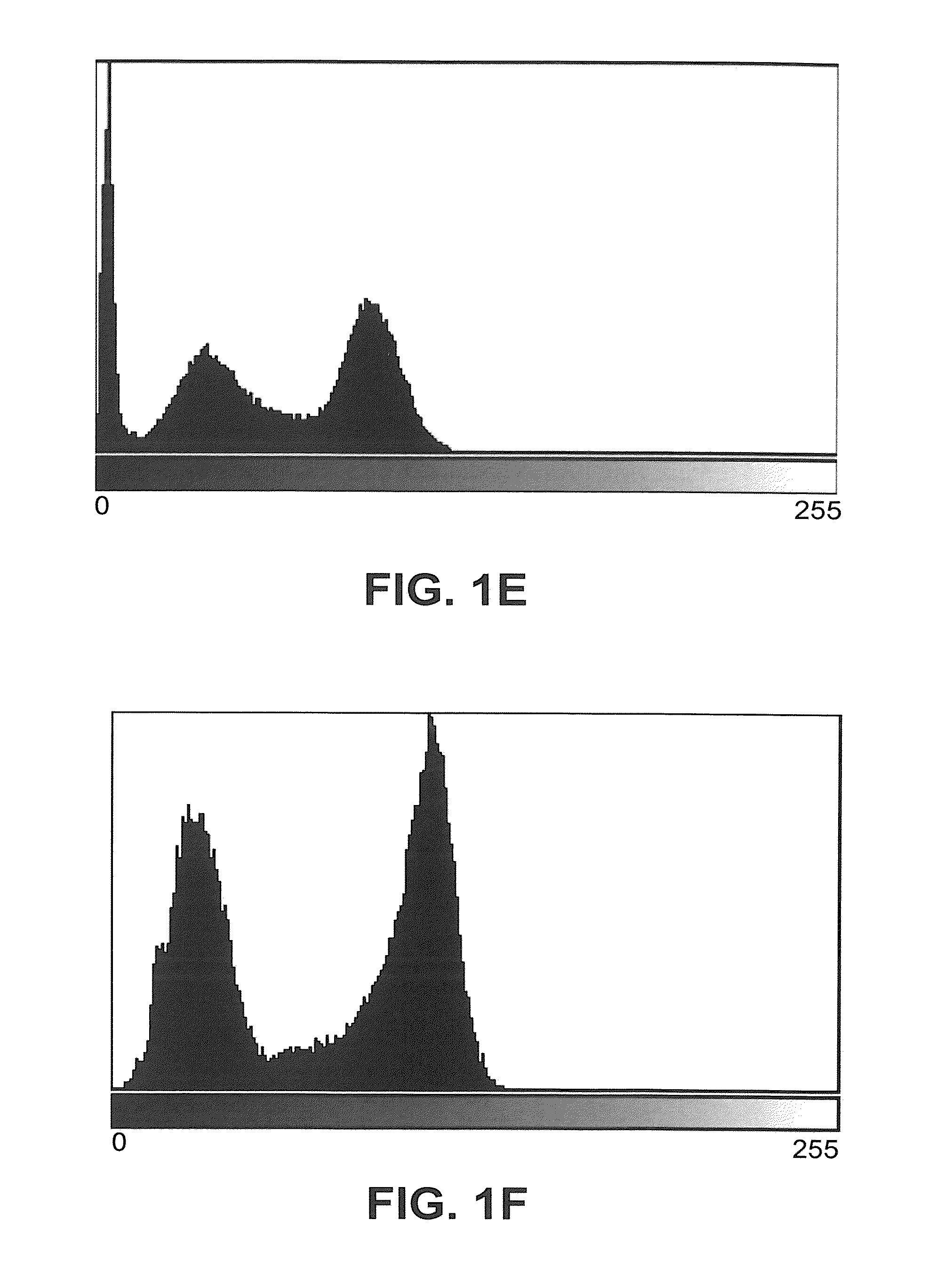
[0052]Image acquisition. Images useable with the subject invention can be acquired using various techniques and equipment. In one embodiment, centrosomal images were acquired in the Analytic Microscopy Core at the H. Lee Moffitt Cancer Center. A549 lung cancer cells and BEAS 2B normal bronchial epithelial cells were grown in RPMI with 10% FBS and BEGM supplemented with BEGM bullet kit, respectively. Cells were plated and grown on coverslips in a 6-well plate at 37° C. with 5% CO2. Cells were fixed using 4% Paraformaldehyde solution for 30 min at 4° C. and permeabilized using 0.5% Triton X solution. Following blocking with 2% BSA, cells were stained with γ-Tubulin antibody (Sigma). Cells were then incubated with AlexaFluor 594 secondary antibody and mounted using ProLong Antifade with DAPI (Invitrogen). A DMI6000 inverted Leica TCS AOBS SP5 tandem-scanni...

example 2

Discriminant and Prognosis of Stage I Long-Term and Short-Term Survival Lung Cancer Patients through Quantitative Analysis of Centrosomal Features

[0110]Tissue processing and immunohistochemisty

[0111]35 cases of lung tissue with different forms and stage of cancer were provided from H. Lee Moffitt Cancer Center & Research Institute. Samples were processed within the Moffitt Microarray Core. Tissues were fixed in formaldehyde, and then embedded in paraffin. For immunohistochemistry, sections were deparafinized using xylene and ethanol washes followed by antigen retrieval buffer (Dako). According to protocol, sections were washed in PBS then blocked (10% normal goat serum, 3% BSA and 0.5% gelatin in PBS) for 1 hour at 4C. Sections were incubated overnight at 4C in primary antibody anti-gamma Tubulin, produced in rabbit (Sigma, 1:300 diluted in PBS with 2% Normal Goat Serum. Sections were washed in PBS-T, incubated with secondary antibody AlexaFlour 633 goat anti-rabbit IgG (Invitrogen,...


PUM


Abstract

The subject invention concerns methods for the detection, diagnosis, and/or prognosis of cancer by analyzing centrosomal features. In one embodiment, a method includes receiving an image of one or more cells; selecting a region of interest in one cell; segmenting the region of interest to delineate at least one centrosomal; extracting one or more features from the segmented image; and analyzing the extracted features to diagnose cancer. In another embodiment, the progression of cancer can be predicted through analysis and classification of the extracted features. In one embodiment, the method can be performed by a quantitative cancer analysis system including a diagnosis module and/or a prognosis module. In one embodiment, the method can be performed using an image processing system.

Description

CROSS-REFERENCE TO RELATED APPLICATION[0001]The present application claims the benefit of U.S. Provisional Application Ser. No. 61 / 225,003, filed Jul. 13, 2009, which is hereby incorporated by reference herein in its entirety, including any figures, tables, or drawings.FIELD OF INVENTION[0002]This invention relates to the detection, diagnosis, and / or prognosis of cancers. Specifically, the invention provides an imaging algorithm to analyze centrosomal features in order to distinguish cancer cells from normal cells and prognose long term from short term survival cancer patients.BACKGROUND OF INVENTION[0003]The centrosome is a cellular organelle that functions as the microtubule organizing center of interphase and mitotic cells (Nigg (2002)). The centrosome duplicates itself only once during each cell cycle with duplication beginning near the G1-S transition and completing during the G2 phase. Duplicated centrosomes separate to produce two mitotic spindle poles that organize the mitot...

Claims


Application Information

Patent Timeline
IPC IPC(8): G06K9/00
CPCA61B10/02G01N33/5076G01N2800/52G06T7/0012G06K9/0014G01N33/574G06V20/695
Inventor ZHUKOV, TATYANA A.SONG, DANSHENGTOCKMAN, MELVYN S.
Owner H LEE MOFFITT CANCER CENT & RES INST INC
Why Eureka
Try Eureka