

Processing of abdominal images
a processing and image technology, applied in the field of abdominal image processing, can solve the problems of reducing the interaction time needed, and reducing the number of failures
- Summary
- Abstract
- Description
- Claims
- Application Information
AI Technical Summary
Problems solved by technology
Method used
Image

Examples
Embodiment Construction
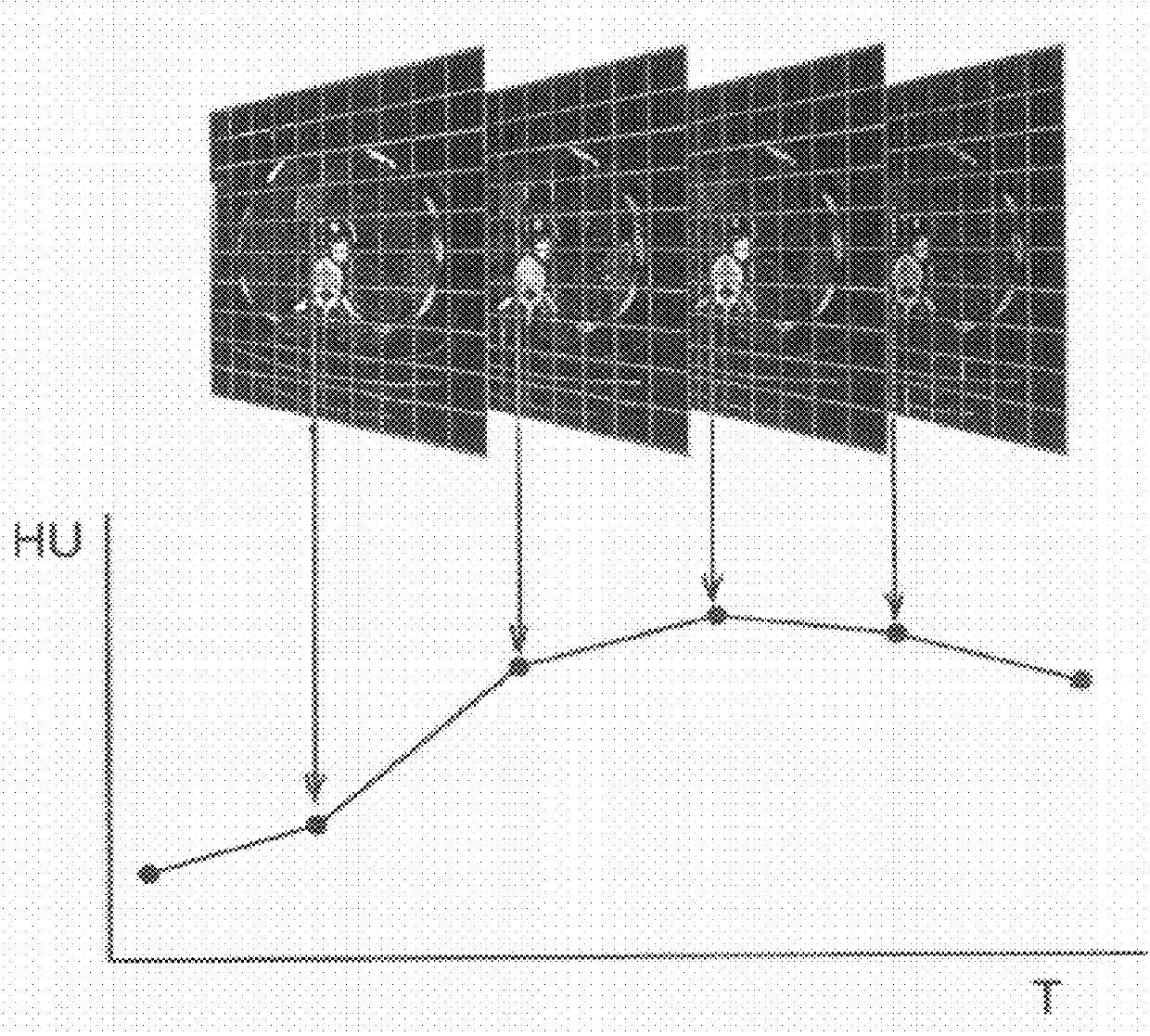
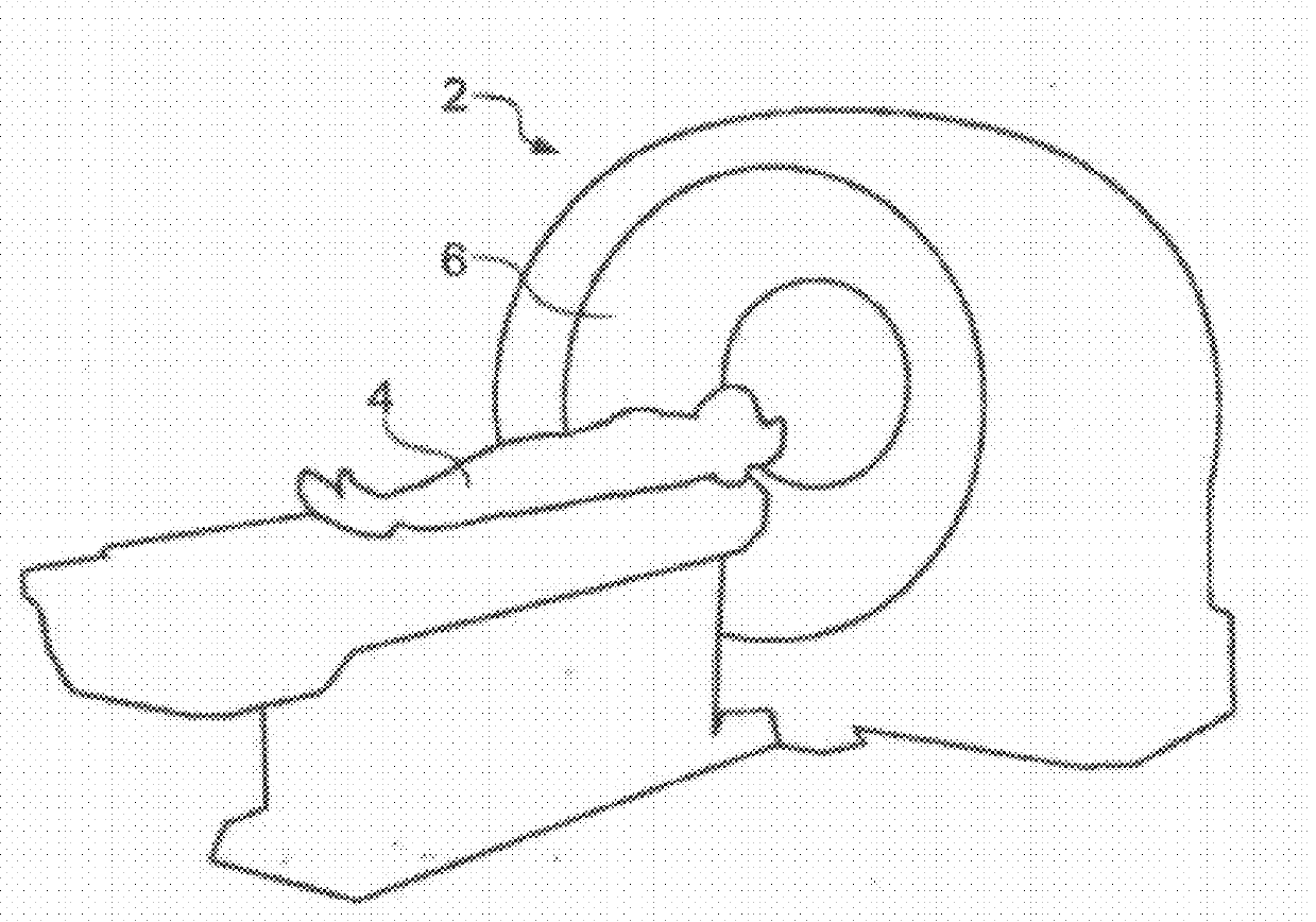
[0018]FIG. 2 is a schematic perspective view of a generic scanner 2, most especially a computer-assisted tomography (CT) scanner, for obtaining a 3D X-ray scan of a region of a patient 4. A patient's abdomen including one or more organs or other anatomical features of interest is placed within a circular opening 6 of the scanner 2. A series of image slices through the patient's abdomen is taken. Raw image data are derived from the scanner and could comprise a collection of one thousand 2D 512×512 data subsets, for example. These data subsets, each representing a slice of the region of the patient being studied, are combined to produce volume data. The volume data, which makes up a 3D image data set, comprise a collection of voxels each of which corresponds to a pixel in one of the slices. Thus the volume data are a 3D representation of the feature imaged and various user-selected 2D projections (output images) of the 3D representation can be displayed (typically on a computer monito...
PUM
 Abstract
Description
Claims
Application Information