Supercharge Your Innovation With Domain-Expert AI Agents!

Ultrasound diagnostic apparatus and method of displaying medical image thereof

a diagnostic apparatus and ultrasound technology, applied in the field of ultrasound diagnostic equipment, can solve the problems of displaying the same cross-section of the reference medical image and the ultrasound image in a longer time, and unable to grasp which section of the subject a reference medical image, and achieve the effect of convenient alignmen

Active Publication Date: 2012-12-06
GENERAL ELECTRIC CO
View PDF3 Cites 16 Cited by
  • Summary
  • Abstract
  • Description
  • Claims
  • Application Information

AI Technical Summary

Benefits of technology

[0012]When an operator inputs a command for displaying a reference medical image, including a characteristic region, in which the location on a subject is identified, a cross-section of the reference medical image thereof is displayed. Since the cross-section of the reference medical image is displayed in which the relative location on subject is identified, a cross-section of a real-time ultrasound image having the same cross-section as the reference medical image can be easily displayed. Therefore, the location can be easily aligned.
[0013]Also, the cross-section in which the reference medical image is displayed in relation to the subject can be easily identified by referring to the marker.
[0014]Further, a portion of the cross-section of the reference medical image in relation to the subject can be easily identified by referring to the displayed distance.

Problems solved by technology

When the ultrasound diagnostic apparatus operator who is unaccustomed to the X-ray CT image or the MRI image performs such location alignment, one may not be able to grasp which section in the subject a reference medical image is even if the reference medical image is displayed.
In some cases, finding a cross-section of the reference medical image having a characteristic region for location alignment takes a longer time, and displaying the same cross-sections of the reference medical image and the ultrasound image takes a longer time.

Method used


Image

  • Ultrasound diagnostic apparatus and method of displaying medical image thereof
  • Ultrasound diagnostic apparatus and method of displaying medical image thereof
  • Ultrasound diagnostic apparatus and method of displaying medical image thereof

Examples


Embodiment Construction

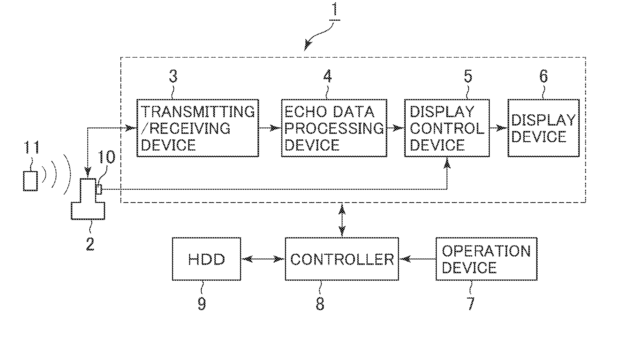
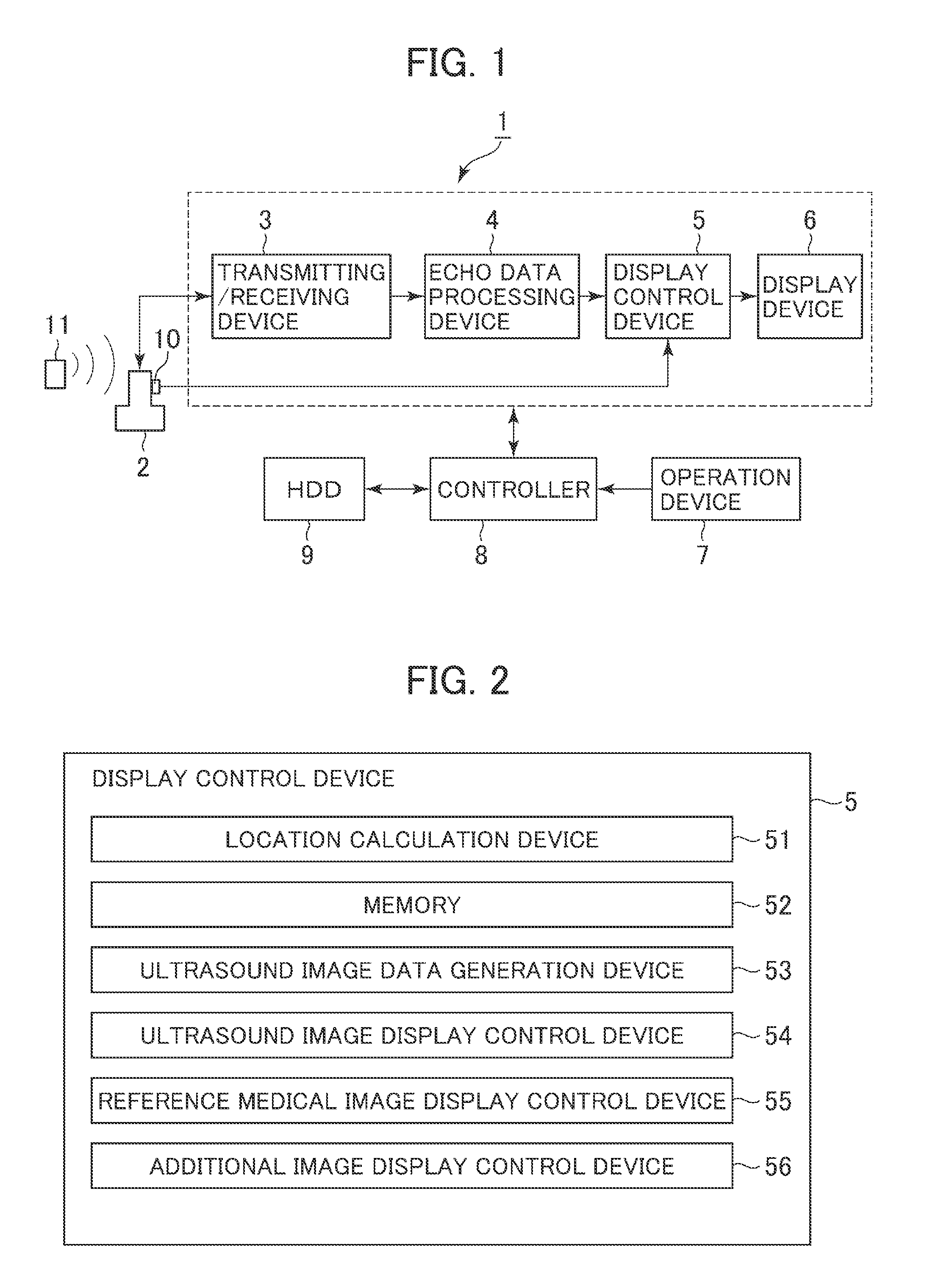
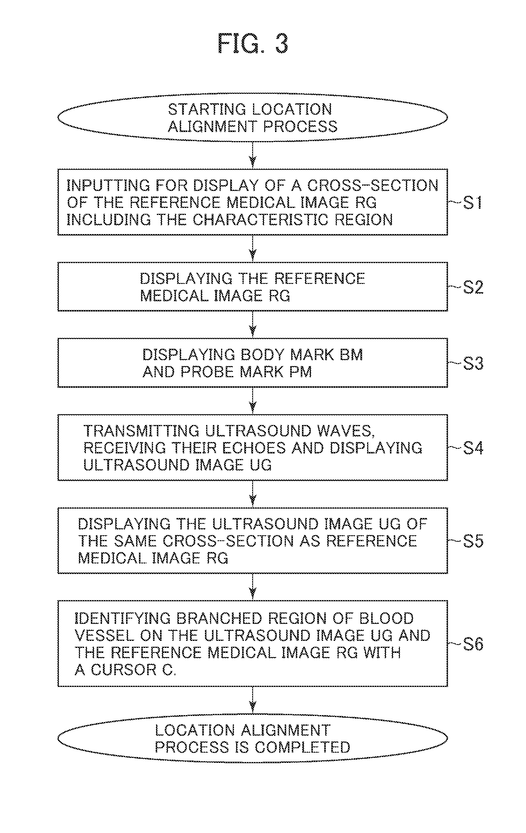
[0028]Hereinafter, exemplary embodiments are explained with reference to FIG. 1 through FIG. 8. An ultrasound diagnostic apparatus 1 shown in FIG. 1 includes an ultrasound probe 2, a transmitting / receiving device 3, an echo data processing device 4, a display control device 5, a display device 6, an operation device 7, a controller 8 and a HDD (Hard Disk Drive) 9.

[0029]The ultrasound probe 2 is configured with an array of ultrasound transducers (not shown), and the ultrasound transducers transmit ultrasound waves to a subject and receive their echo signals.

[0030]The magnetic sensor 10 includes Hall elements and is provided on the ultrasound probe 2, for example. Magnetic field generated from the magnetic field generation device 11 including a magnetic field generation coil is detected by the magnetic sensor 10. The signal detected at the magnetic sensor 10 is inputted to the display control device 5. The signal detected at the magnetic sensor 10 can be inputted to the display contro...


PUM


Abstract

An ultrasound diagnostic apparatus is provided. The ultrasound diagnostic apparatus includes an input device configured to receive an input to display a cross-section of a reference medical image, wherein the reference medical image identifies a location of the cross-section in relation to a subject, and includes a characteristic region, and a reference medical image display control device configured to display the cross-section of the reference medical image based on volume data of the reference medical image previously acquired, the cross-section displayed according to the input.

Description

CROSS REFERENCE TO RELATED APPLICATIONS[0001]This application claims the benefit of Japanese Patent Application No. 2011-120467 filed May 30, 2011, which is hereby incorporated by reference in its entirety.BACKGROUND OF THE INVENTION[0002]The present invention relates to an ultrasound diagnostic apparatus displaying an ultrasound image and a reference medical image. The present invention also relates to a method of displaying a medical image thereof.[0003]A conventional ultrasound diagnostic apparatus can display a real-time ultrasound image based on an echo signal acquired by transmitting an ultrasound wave from an ultrasound probe. For example, WO2004-098414 discloses an ultrasound diagnostic apparatus which displays a real-time ultrasound image and a reference medical image such as an X-ray CT (Computed Tomography) image and an MRI (Magnetic Resonance Imaging) image of the same cross-section in relation to a subject.[0004]This ultrasound diagnostic apparatus identifies a region c...

Claims


Application Information

Patent Timeline
IPC IPC(8): A61B8/13
CPCA61B8/00A61B8/463A61B8/5261A61B8/466A61B8/483A61B8/464
Inventor YAWATA, TSUTOMU
Owner GENERAL ELECTRIC CO
Features
  • R&D
  • Intellectual Property
  • Life Sciences
  • Materials
  • Tech Scout
  • Unparalleled Data Quality
  • Higher Quality Content
  • 60% Fewer Hallucinations
Learn More