Method for Compensating for Respiratory Motion in Magnetic Resonance Imaging

a magnetic resonance imaging and respiratory motion technology, applied in the field of motion compensation in magnetic resonance imaging, can solve the problems of imposing a limit on the speed at which imaging can take place, requiring appreciably more time to obtain a high resolution mr image of the heart, and a particular problem of respiration-induced motion of the hear

Inactive Publication Date: 2012-12-20
UNIVERSITY OF CAPE TOWN
View PDF3 Cites 11 Cited by
  • Summary
  • Abstract
  • Description
  • Claims
  • Application Information

AI Technical Summary

Benefits of technology

[0009]It is an object of this invention to provide a method for compensating for respiratory motion in magnetic resonance imaging which

Problems solved by technology

The inherent relaxation time of the tissue being imaged, however, imposes a limit on the speed at which imaging can take place.
Obtaining a high resolution MR image of the heart therefore requires appreciably more time than imaging other soft tissue.
Respiration-induced motion of the heart is a particular problem for high resolution cardiac MR imaging.
As the imaging spans several heartbeats, the subject's breathing can cause blurring of the acquired image due to the displacement of the heart within the chest cavity as the subject inhales and exha

Method used


Image

  • Method for Compensating for Respiratory Motion in Magnetic Resonance Imaging
  • Method for Compensating for Respiratory Motion in Magnetic Resonance Imaging
  • Method for Compensating for Respiratory Motion in Magnetic Resonance Imaging

Examples


Embodiment Construction

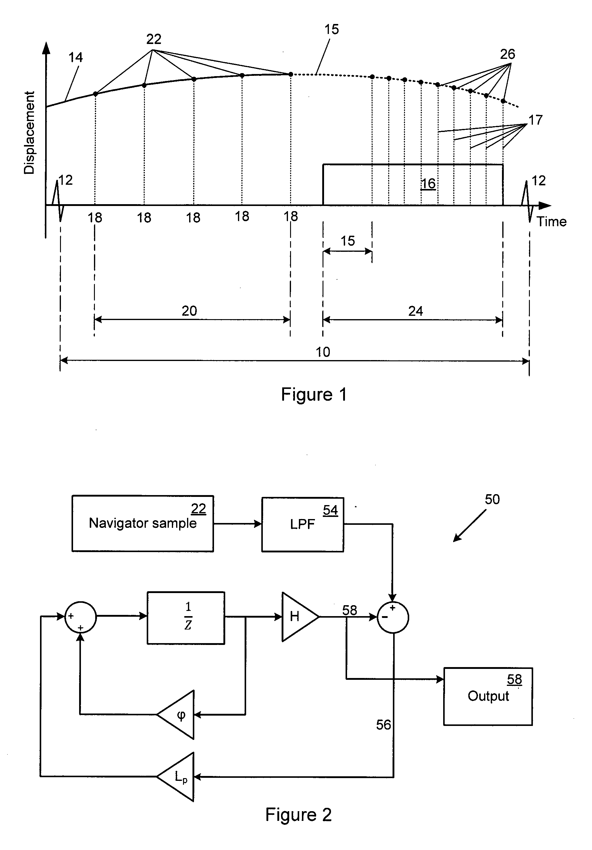
[0049]FIG. 1 is a graph illustrating a method for compensating for respiration-induced motion of a subject's heart as it is imaged by a magnetic resonance (MR) scanner. The horizontal axis represents time and the graph is shown over one cardiac cycle (10) which spans a pair of consecutive heartbeats (12). The vertical axis represents the displacement of the subject's heart that occurs due to the subject breathing.

[0050]Image preparation and acquisition by the MR scanner is done during a segment (16) which is gated by an ECG trigger signal which detects the heartbeats (12) so as to occur at the same time during consecutive cardiac cycles. For a healthy subject with a resting heart rate of about 70 beats per minutes, the imaging segment (16) can be about 200 ms long, which includes a scanner preparation period (15) followed by an imaging sequence during which about 25 image projection views or lines (17) can be obtained during the single cardiac cycle.

[0051]The movement of the subject...


PUM


Abstract

A method for compensating for respiratory motion of a structure imaged by a magnetic resonance (MR) scanner, such as heart, is disclosed. The method comprises obtaining a sequence of navigator samples (22) representative of the progressive displacement of the structure during a first time interval (20), and adjusting a waveform (14) of predefined initial parameters to follow the sequence of navigator samples. During a second time interval (24) which follows the first time interval and during which the magnetic resonance scanner takes a number of image projection views or lines (17), the expected respiratory displacement of the structure is estimated at the times that the image projection lines are taken, by extrapolating the waveform model and estimating the displacements (26) predicted by the extrapolated waveform, so that the position at which the image projection lines are taken can be adjusted to compensate for the respiratory motion, so as to yield a sharper magnetic resonance image.

Description

FIELD OF THE INVENTION[0001]This invention relates to motion compensation in magnetic resonance imaging and, more specifically, to a method for compensating for respiratory motion in magnetic resonance imaging of structures affected by such motion, such as structures within a chest cavity of a subject, and especially structures within the chest cavity also affected by other cyclic or regular motion, such as the heart.BACKGROUND TO THE INVENTION[0002]Magnetic resonance imaging (MRI) relies on the excitation and relaxation of sub-atomic particles within the structure being imaged, and is an important technology for imaging soft tissue in humans and animals. The inherent relaxation time of the tissue being imaged, however, imposes a limit on the speed at which imaging can take place. Typically, each 2D image (called a “slice”) is built up from several hundred image projection views or lines, which (depending on the required resolution) can each take between 3 to 6 milliseconds to compl...

Claims


Application Information

Patent Timeline
IPC IPC(8): G01R33/563
CPCG01R33/5676
Inventor BURGER, IAN HAMILTON
Owner UNIVERSITY OF CAPE TOWN
Why Eureka
Try Eureka