Interventional radiologic devices and methods for embedded filter removal

a radiologic device and filter technology, applied in medical science, surgery, diagnostics, etc., can solve the problems of not being able to retrieve the filter, the filter may be permanently embedded in the ivc, and cannot be removed using basic, so as to reduce the risks of complex filter removal, safe and effective removal of embedded ivc filters, and minimal blood loss

Inactive Publication Date: 2013-02-28
THE BOARD OF TRUSTEES OF THE LELAND STANFORD JUNIOR UNIV
View PDF6 Cites 1 Cited by
  • Summary
  • Abstract
  • Description
  • Claims
  • Application Information

AI Technical Summary

Benefits of technology

[0009]Embodiments of the invention has several advantages. For example, the device can be inserted over 2 wires simultaneously thereby allowing insertion over a wire loop configuration used to engage the apex of embedded IVC filters. The strength of the sheath material is custom tailored to withstand the forces necessary to remove an embedded IVC filter. The material is also puncture resistant to any sharp edges of the captured filter during removal through the sheath. Modular force meter attachments can be used to allow constant monitoring of tension applied to the embedded filter to allow operation within a safe window of tension during complex filter retrieval, thereby reducing the risks associated with complex filter removal. A special valve system (for example, but not limited to, a valve with an inflatable balloon-type diaphragm made of puncture resistant material) is designed to accommodate multiple device components simultaneously when used to remove the filter, while providing hemostasis. In addition, a dual-valve assembly option with containment chamber for the filter allows facile delivery of the filter with minimal blood loss. The sheath tip and construction is designed specifically for embedded filter removal using a proper angle and shape. The complete device and apparatus is specifically designed to allow safe and effective removal of embedded IVC filters.

Problems solved by technology

Although standard techniques for removal of inferior vena cava (IVC) filters are successful in most cases, an estimated 20-40%, of retrievable IVC filters cannot be removed using basic methods and standard apparatus even when attempted within routine retrieval windows.
Additionally, filter retrieval is not attempted in many patients with retrievable filters that have been embedded for a prolonged time and in most patients with permanent filter types, which are by definition considered irretrievable.
All of these filters may intentionally or unintentionally become permanently embedded within the IVC.
A few reports have described the risky endeavor of attempting to separate densely adherent filters from the underlying caval wall using basic apparatus, but there are no current devices specifically designed to remove embedded IVC filters.
Additionally, many patients have a permanent filter type that was never designed for removal.
In all patients with embedded filters, retrieval is not currently considered a feasible routine option for filter management.
Consequently, these patients are subject to all the risks of prolonged IVC filter implantation, including the potential need for lifelong anticoagulation to reduce thrombotic risks related to prolonged filter implantation.

Method used


Image

  • Interventional radiologic devices and methods for embedded filter removal
  • Interventional radiologic devices and methods for embedded filter removal
  • Interventional radiologic devices and methods for embedded filter removal

Examples


Embodiment Construction

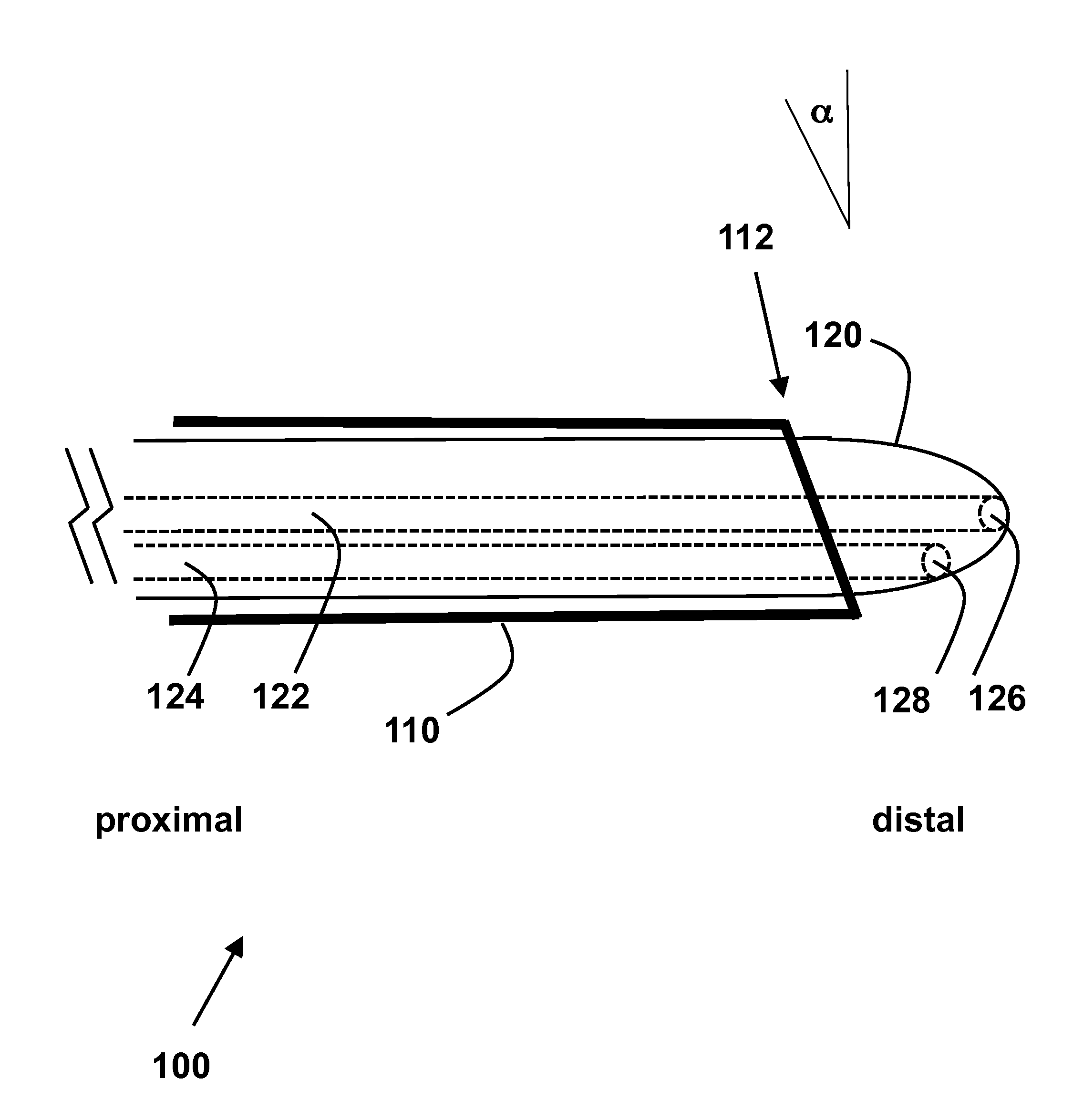
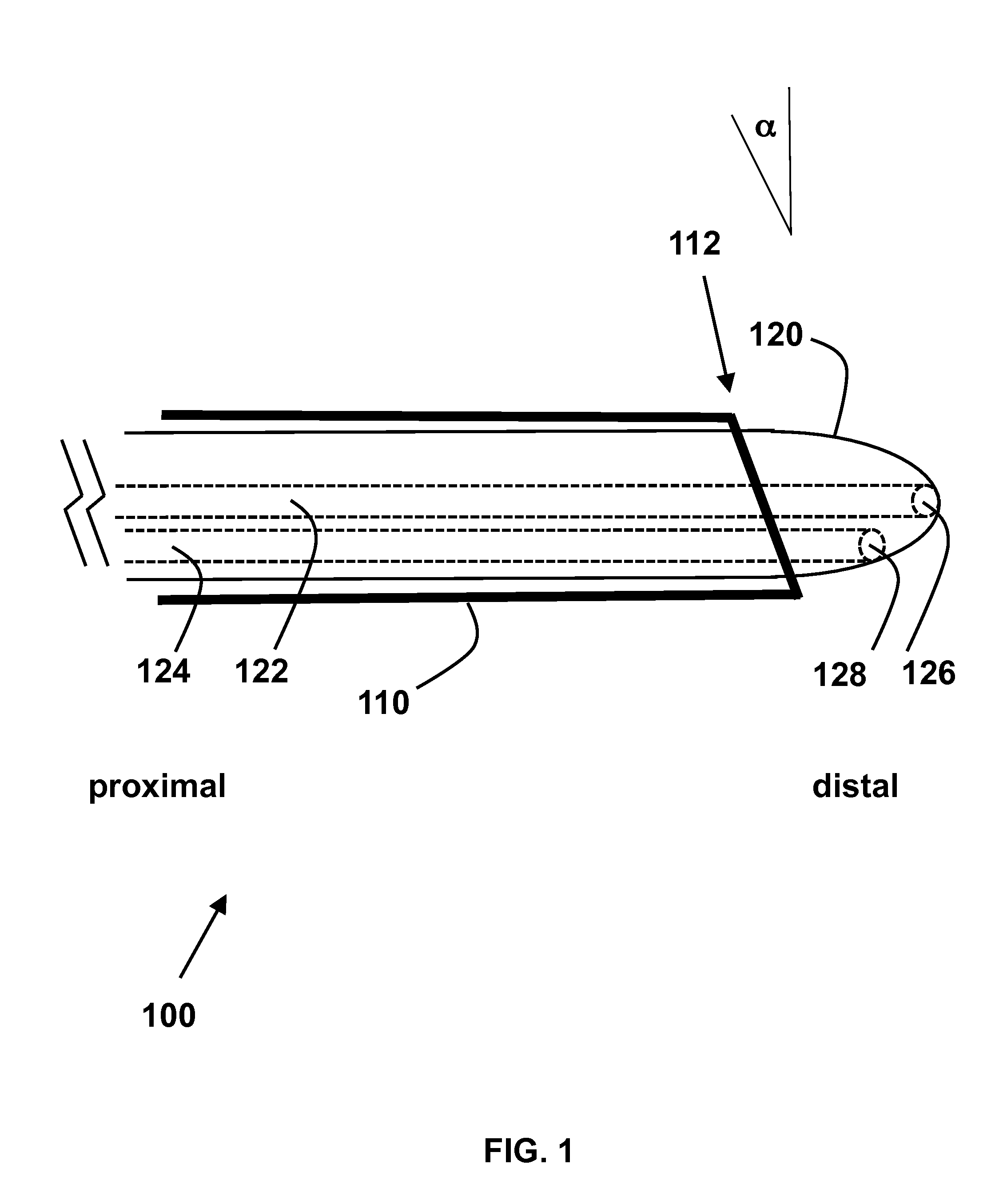
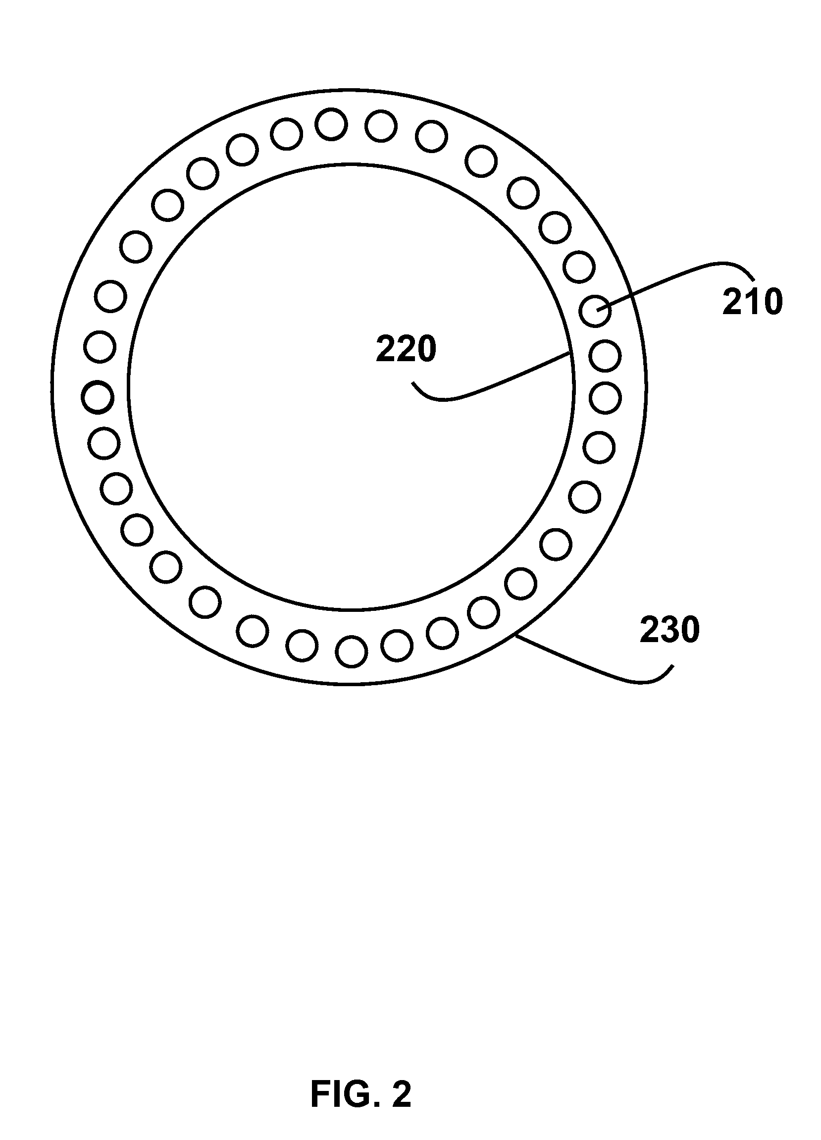
[0018]In one embodiment, the interventional radiologic device for removal of an embedded filter (for example, but not limited to, an embedded inferior vena cava filter) includes a laser sheath, one or more modular adapters, and a removable introducer. As shown by device 100 in FIG. 1, the laser sheath 110 has an inner diameter (i.e. inner wall) surrounding a lumen between a proximal open end and a distal open end. In one example, the distal open end is beveled having an angle up to 45 degrees (bevel is indicated by 112 and measures as angle α). In one example, the laser sheath is made of reinforced polymer sheath material. In another example, the laser sheath is made of reinforced polymer sheath material which is able to withstand up to 10 pounds of pressure without getting fractured. In yet another example, puncture resistant materials could be used. Through the wall of the laser sheath (220 and 230 are respectively the inner wall and outer wall of the laser sheath), optical fibers...


PUM


Abstract

An interventional radiologic device for removal of an embedded filter is provided. The device distinguishes a laser sheath with optical fibers for applying ablation energy to tissue. A removable introducer is movable within the lumen of the laser sheath and has at least two open channels to run wires. One or more sheath detachable modular adapters are used for providing ablation energy, force sensing and/or preventing bleeding. The distal open end of the laser sheath is preferably beveled with an angle up to 45 degrees. In one aspect, the introducer has a bulb-like tip, which fits against the distal end the laser sheath, yet can still be removed from the laser sheath via its proximal end. This device can be inserted over two wires simultaneously thereby allowing insertion over a wire loop configuration used to engage the apex of embedded IVC filters.

Description

CROSS-REFERENCE TO RELATED APPLICATIONS[0001]This application claims priority from U.S. Provisional Patent Application 61 / 575,521 filed Aug. 22, 2011, which is incorporated herein by reference.FIELD OF THE INVENTION[0002]This invention relates to surgical devices and medical procedures. In particular, the invention relates to interventional radiologic devices and methods for removing embedded filters.BACKGROUND OF THE INVENTION[0003]Although standard techniques for removal of inferior vena cava (IVC) filters are successful in most cases, an estimated 20-40%, of retrievable IVC filters cannot be removed using basic methods and standard apparatus even when attempted within routine retrieval windows. Additionally, filter retrieval is not attempted in many patients with retrievable filters that have been embedded for a prolonged time and in most patients with permanent filter types, which are by definition considered irretrievable. All of these filters may intentionally or unintentional...

Claims


Application Information

Patent Timeline
Patent Type & Authority Applications(United States)
IPC IPC(8): A61B18/24
CPCA61B18/24A61B2019/464A61B2018/208A61B2018/00172A61B2090/064
Inventor KUO, WILLIAM T.
Owner THE BOARD OF TRUSTEES OF THE LELAND STANFORD JUNIOR UNIV
Why Eureka
Try Eureka