System and method for controlling calibration and delay phases of parallel, contrast-enhanced magnetic resonance imaging

a technology of contrast enhancement and imaging, applied in the field of magnetic resonance imaging, can solve the problems of insufficient acquisition time, overhead time required for parallel acquisition, and inability to achieve sufficient acquisition time, so as to improve image quality, reduce the delay associated with pulse sequence download, and simplify the preparation and examination process

Active Publication Date: 2013-03-14
MAYO FOUND FOR MEDICAL EDUCATION & RES
View PDF4 Cites 9 Cited by
  • Summary
  • Abstract
  • Description
  • Claims
  • Application Information

AI Technical Summary

Benefits of technology

[0014]The present invention overcomes the aforementioned drawbacks by providing a method for parallel imaging in which calibration data is acquired during the time period when the contrast agent is in transit from the injection site to the targeted arterial vasculature. By doing so, the preparation and examination process can be greatly streamlined and the delays associated with pulse sequence downloads eliminated. Furthermore, image quality, for example, in abdominal magnetic resonance angiography studies, can be improved by allowing all data acquisition to be done within a single breathhold. Thus, the present invention improves the temporal scan requirements of parallel imaging acquisitions.

Problems solved by technology

There are technical challenges associated with performing CE-MRA.
First, to obtain a three-dimensional (3D) image with adequate spatial resolution, it is necessary to have a sufficiently long acquisition time.
The key point of this discussion is that for both approaches to parallel imaging, there is overhead time required for the implementation of the parallel acquisition, primarily due to the need to acquire calibration data.
Although this may not, in the abstract, seem like a long duration, it can be highly problematic for imaging of the thoracic, abdominal, or pelvic vasculature.
If this is not the case, then the mismatch between object positions can cause ghost-like artifacts which can interfere with interpretation of the contrast-enhanced images.
Thus, this duration can be even further extended, which undermines the speed and efficiency sought to be gained by the use of parallel imaging.

Method used


Image

  • System and method for controlling calibration and delay phases of parallel, contrast-enhanced magnetic resonance imaging
  • System and method for controlling calibration and delay phases of parallel, contrast-enhanced magnetic resonance imaging
  • System and method for controlling calibration and delay phases of parallel, contrast-enhanced magnetic resonance imaging

Examples


Embodiment Construction

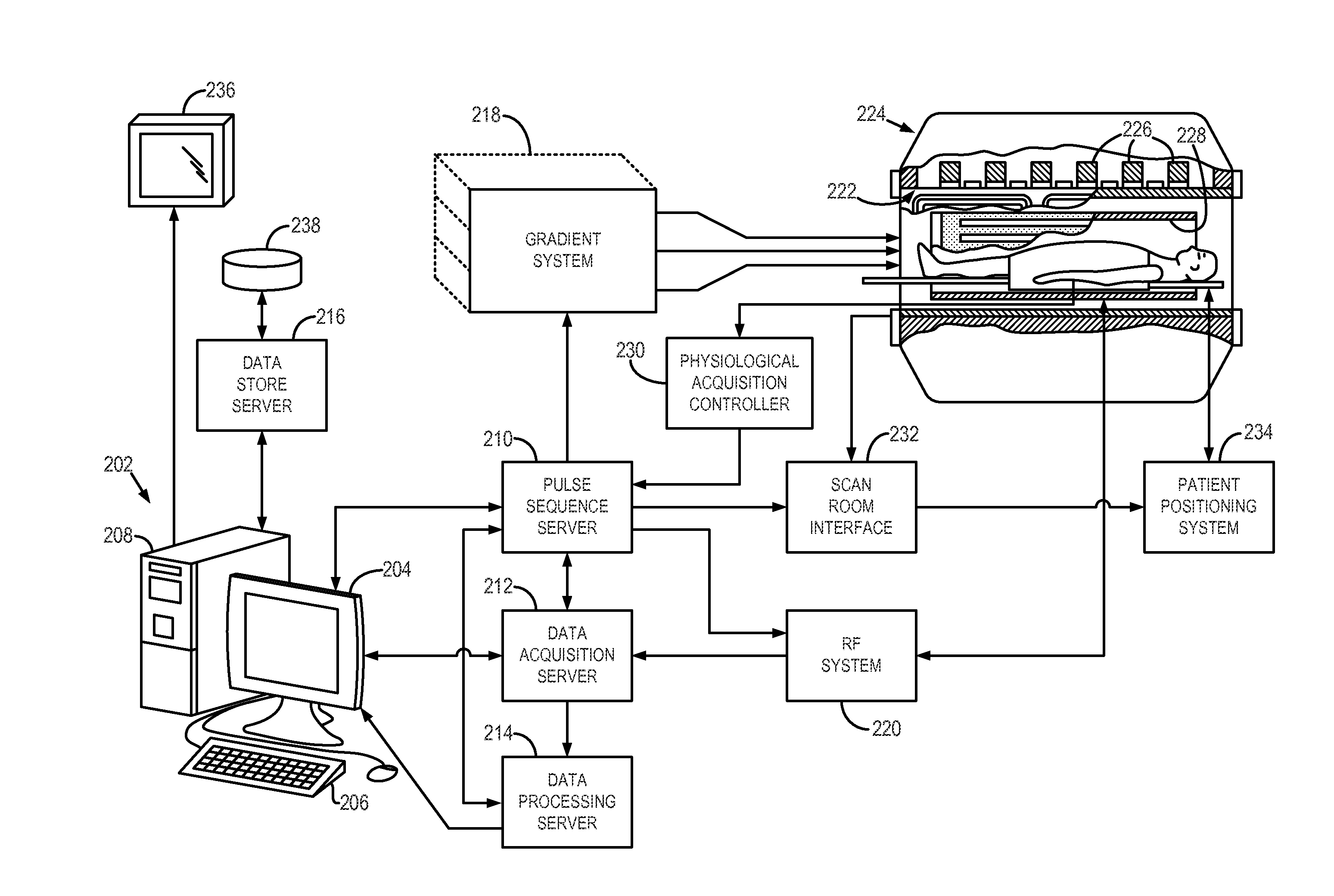
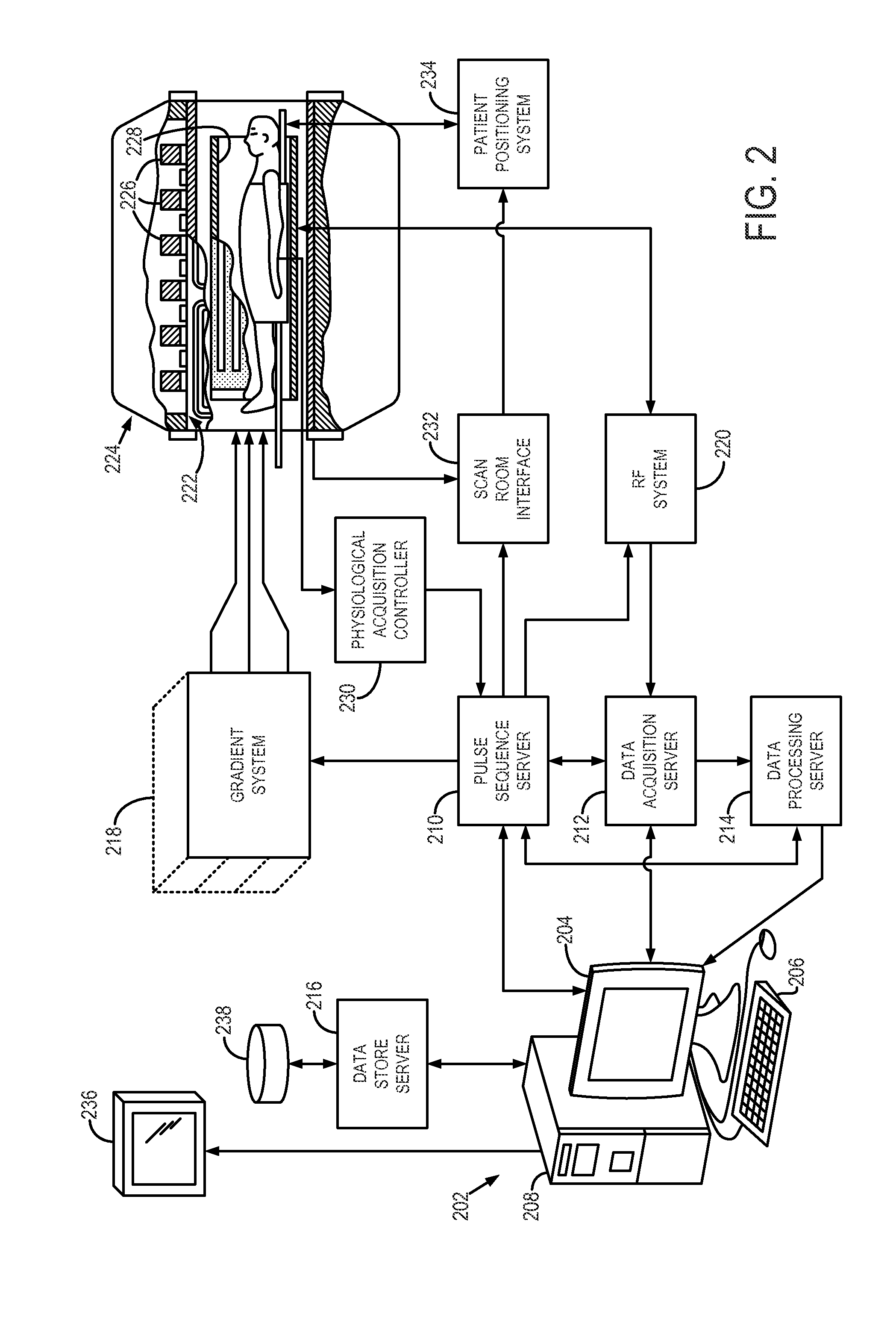
[0027]Referring particularly now to FIG. 2, an exemplary magnetic resonance imaging (“MRI”) system 200 capable of practicing embodiments of the present invention is illustrated. The MRI system 200 includes a workstation 202 having a display 204 and a keyboard 206. The workstation 202 includes a processor 208, such as a commercially available programmable machine running a commercially available operating system. The workstation 202 provides the operator interface that enables scan prescriptions to be entered into the MRI system 200. The workstation 202 is coupled to four servers: a pulse sequence server 210; a data acquisition server 212; a data processing server 214, and a data store server 216. The workstation 202 and each server 210, 212, 214, and 216 are connected to communicate with each other.

[0028]The pulse sequence server 210 functions in response to instructions downloaded from the workstation 202 to operate a gradient system 218 and a radiofrequency (“RF”) system 220. Grad...


PUM


Abstract

A system and method for performing parallel magnetic resonance angiography includes controlling operation of a magnetic gradient system and an RF system to perform a calibration data pulse sequence to begin acquiring calibration data for use in a parallel imaging reconstruction process after receiving an indication that the subject has received a dose of a contrast agent. The acquisition of the calibration data is discontinued before the contrast agent reaches a peak concentration within a region of interest (ROI) of the subject and operation of the magnetic gradient system and RF system is controlled to perform an imaging pulse sequence in accordance with a parallel imaging acquisition to begin acquiring image data from the ROI. The image data is reconstructed into an image of the ROI using the calibration data.

Description

CROSS-REFERENCE[0001]This application is based on, claims priority to, and incorporates herein by reference in its entirety, U.S. Provisional Application Ser. No. 61 / 532,487, entitled, “Modified Calibration of SENSE-Accelerated Time-Resolved Contrast Enhanced MRA,” and filed Sep. 8, 2011.STATEMENT REGARDING FEDERALLY SPONSORED RESEARCH[0002]This invention was made with government support under HL070620 awarded by the National Institutes of Health. The government has certain rights in the invention.BACKGROUND OF THE INVENTION[0003]The present invention relates generally to systems and methods for magnetic resonance imaging (“MRI”) and, more particularly, the invention relates to systems and methods for image acquisition of contrast-enhanced magnetic resonance angiography images using parallel-imaging MRI techniques.[0004]Contrast-enhanced magnetic resonance angiography (“CE-MRA”) is a method whereby magnetic resonance imaging (“MRI”) techniques are used to image blood vessels of the ...

Claims


Application Information

Patent Timeline
Patent Type & Authority Applications(United States)
IPC IPC(8): G01R33/48
CPCG01R33/5635G01R33/5611A61B5/05G01R33/20G01R33/285G01R33/483G01R33/54G01R33/56
Inventor RIEDERER, STEPHEN J.
Owner MAYO FOUND FOR MEDICAL EDUCATION & RES
Why Eureka
Try Eureka