Method of determination of access areas from 3D patient images

a 3d patient and access area technology, applied in image analysis, medical science, surgery, etc., can solve the problems of difficult to determine the correct and full diagnosis of this pathology, the repetitive impact of the proximal femoral neck, and the complex structure of the human body

Active Publication Date: 2013-04-18
SMITH & NEPHEW ORTHOPAEDICS
View PDF3 Cites 57 Cited by
  • Summary
  • Abstract
  • Description
  • Claims
  • Application Information

AI Technical Summary

Benefits of technology

[0027]According to another embodiment, the planning information is a projection of an optimal surface to be built onto the bone.

Problems solved by technology

Articulations of the human body are often very complex systems and no precise generic model exists to capture all the variability from one articulation to another.
Structural abnormalities in the morphology of the hip can limit motion and result in repetitive impact of the proximal femoral neck against the acetabular labrum and its adjacent cartilage.
However, the correct and full diagnosis of this pathology is not easy to determine, especially when dealing with subtle deformities.
The processing of the 3D image remains a laborious manual task which cannot ensure accuracy and reproducibility, potentially misleading the diagnosis or the surgical indication.
Moreover, even though 3D information can be extracted from such exams, the reconstructed bone volumes remain static and cannot predict with reliability the exact location of the impingement which occurs during the mobilization of the hip.
However, due to the depth of the joint and the reduced visibility and access, theses hip arthroscopy procedures are difficult to perform and not all surgeons feel comfortable about adopting the technique.
Failure of arthroscopic procedures for FAI is most commonly associated with incomplete decompression of the bony lesions.
However, in minimally invasive (MIS) procedures such as arthroscopy, the patient anatomy access is generally reduced and cannot allow for a direct digitization of the anatomy.
But in cases such as hip arthroscopy, 2D image information is generally not sufficient to achieve the required accuracy in three dimensions, and a pre-operative 3D image is usually required.

Method used


Image

  • Method of determination of access areas from 3D patient images
  • Method of determination of access areas from 3D patient images
  • Method of determination of access areas from 3D patient images

Examples


Embodiment Construction

:

[0048]The following description is directed to the use of CT images but it can be easily extended to other 3D images such as MR images.

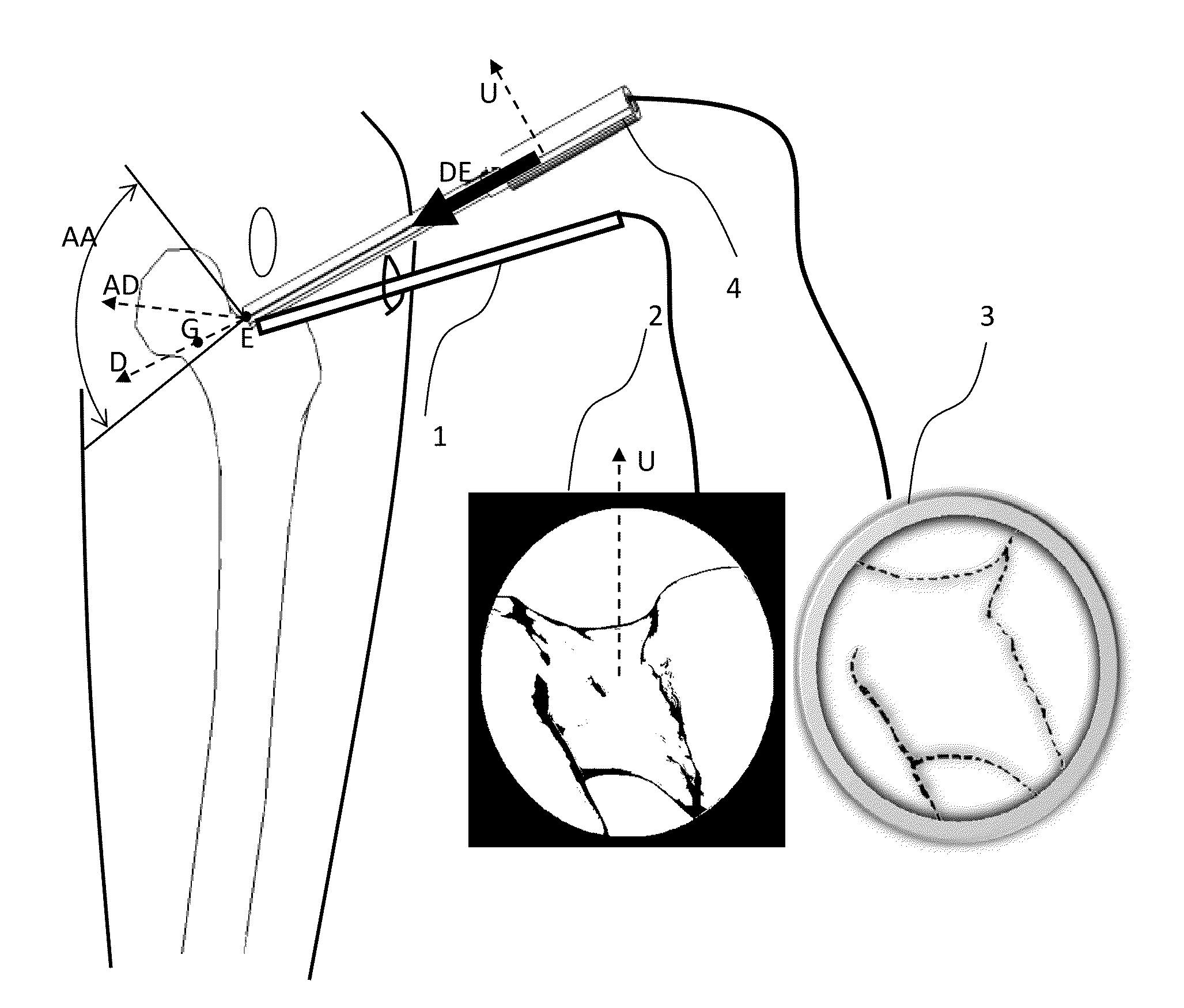
[0049]The purpose of the invention is to provide a method and a device for helping the user to accurately and automatically determine portals and safe access areas for arthroscopic procedures using pre-operative 3D image and also for planning relevant arthroscopic images that can be used during the surgery in addition to target data. It is intended to provide a very simple and fast rehearsal tool or simulation of surgery. Moreover, the invention provides help for the surgeon to locate and use the predefined portals and safe access areas during surgery.

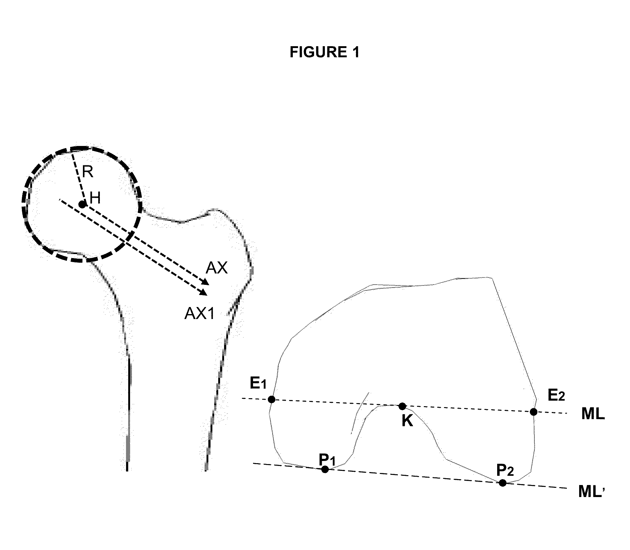
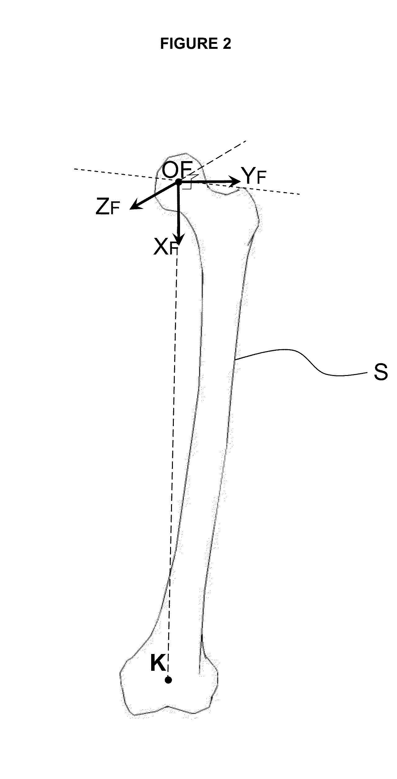
[0050]For clarity, an example of the method of the invention is described for the femur and pelvis bones during a hip arthroscopy procedure in the case of a FAI indication, but it can be applied to any other human or animal bone for which itself or the adjacent ligaments, tendons and muscles present an...


PUM


Abstract

Method of determination of access areas from 3D patient images The invention relates to amethod for automatically determining at least one pre-operative portal for arthroscopy from acquired pre-operative medical images of a bone of a patient, the method comprising the following steps: i)constructing a 3Dsurface(S) of the bone from the 3D image of the bone; ii)determining anatomical landmarks of the bone from the 3D surface; iii)determining a bone reference coordinate system (Rbone); iv)selecting at least one predetermined portal in a database of positions of predetermined portals; v)determining a transform between the bone reference coordinate system (Rbone) and the model coordinate system (Ratlas) so that the bone of the patient and the bone of the reference person are matched in size, in position and/or shape; vi)inferring from the transform and the at least one predetermined portal the pre-operative portal.

Description

TECHNICAL FIELD:[0001]The invention relates to the field of computer assisted surgery, and more particularly to a method for determining portals for arthroscopy.BACKGROUND OF THE INVENTION:[0002]Articulations of the human body are often very complex systems and no precise generic model exists to capture all the variability from one articulation to another. It is therefore necessary to use specific medical images or collection of digital patient data in order to get relevant information to develop techniques, devices and methods that will facilitate a treatment or a diagnosis. Our description focuses on the hip articulation between the acetabulum and the proximal femur although it can be easily extended to other articulations such as shoulder for example.[0003]Structural abnormalities in the morphology of the hip can limit motion and result in repetitive impact of the proximal femoral neck against the acetabular labrum and its adjacent cartilage. Femoro Acetabular Impingement (FAI) i...

Claims


Application Information

Patent Timeline
Patent Type & Authority Applications(United States)
IPC IPC(8): A61B1/317
CPCA61B19/50G06T7/0046A61B1/317G06T2207/30008G06T2207/10072A61B34/10G06T7/75
Inventor CHABANAS, LAURENCELAVALLEE, STÉPHANE
Owner SMITH & NEPHEW ORTHOPAEDICS
Why Eureka
Try Eureka