Magnetic resonance analysis using a plurality of pairs of bipolar gradient pulses
a technology of bipolar gradient pulses and magnetic resonance analysis, applied in the field of magnetic resonance analysis, can solve problems such as signal loss
- Summary
- Abstract
- Description
- Claims
- Application Information
AI Technical Summary
Benefits of technology
Problems solved by technology
Method used
Image
Examples
example 1
Materials and Methods
[0161]Hollow microcapillaries with well defined inner diameter (ID) (PolyMicro Technologies, Phoenix Ariz., USA) were immersed in distilled water for several days prior to each experiment. The water-filled microcapillaries were manually cut to very small pieces (<0.5 cm) and then crushed by mechanical force to “dust”-like small particles. In some cases, the microcapillaries were not crushed but only cut into very small pieces that enabled random orientation within the NMR tube. The resulting porous medium was re-immersed in water for several days, to refill. The porous crush was then poured to an 8 or in some cases 10 mm NMR tube containing Fluorinert (Sigma-Aldrich, Rehovot, Israel). The Fluorinert served, owing to density and polarity differences, to keep any extra-tubular water in the top part of the NMR tube, which was located outside the RF coils.
[0162]The polydisperse specimen was prepared in the same way, only instead of using monodisp...
example 2
Apparent-Susceptibility Induced Anisotropy
[0196]One of the most interesting features of pores embedded within porous media is their local shape and the way these pores are dispersed within the medium. Local pore shape can be considered as spherical or non-spherical (FIGS. 11A and 11B, respectively). However, while there is only one way to disperse spherical pores in a porous medium, locally cylindrical pores, for example, may be either randomly oriented (FIG. 11C) or coherently placed (FIG. 11D) within the porous medium.
[0197]Conventional methods such as s-PFG methods are very limited in depicting the anisotropy, e.g., in the scenario shown in FIG. 11C, since it is extremely difficult to distinguish, for example, spheres from randomly oriented anisotropic pores, as well as to infer on restricted diffusion.
[0198]Many real porous media are heterogeneous in nature and are characterized by background gradients that arise from susceptibility differences within the specimen. The cross-ter...
example 3
Nerve Tissues
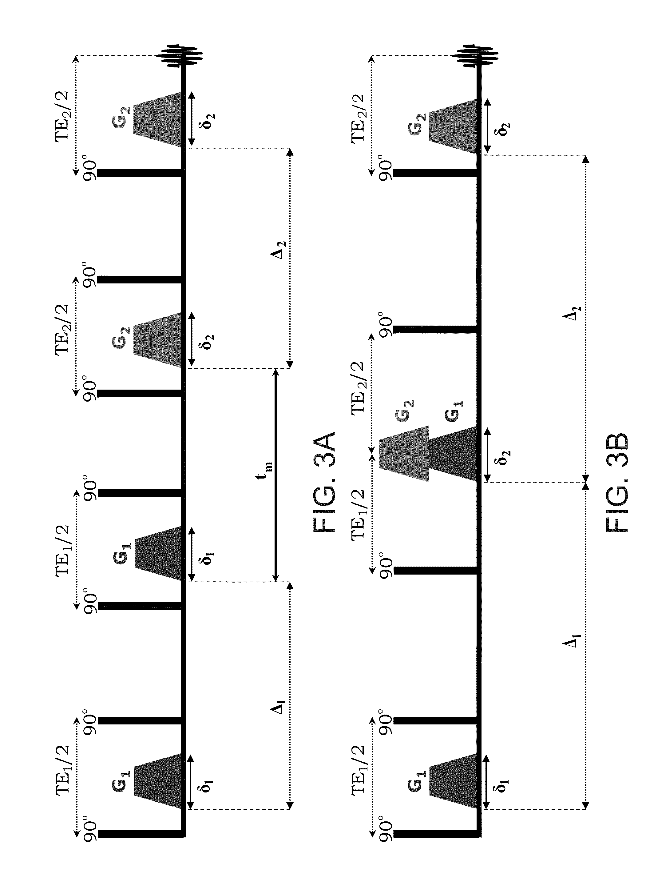
[0230]FIGS. 16A and 16B show E(ψ) profiles obtained from an MR signal received from grey (FIG. 16A) and white (FIG. 16B) matter tissues, in response to the conventional d-PFG sequence shown in FIG. 3B (squares) and the sequence of the present embodiments (circles) shown in FIG. 4C. The experimental parameters for both cases were: 2q=2043 cm−1, tm=0 ms and Δ1=Δ2=50 ms and δ1=δ2=δ3=3 ms.
[0231]For the conventional sequence, the E(ψ) profiles show an erroneous angular dependence both for the grey matter and the white matter tissues.
[0232]On the other hand, the sequence of the present embodiments provided the expected angular dependence for both randomly oriented cells (grey matter) and coherently placed axons (white matter).
[0233]This example demonstrates the ability of the present embodiments to provide microstructural information such as cell shape, size and orientation distribution even in highly heterogeneous biological tissues such as neuronal tissues.
PUM
 Abstract
Description
Claims
Application Information
 - R&D
- Intellectual Property
- Life Sciences
- Materials
- Tech Scout
- Unparalleled Data Quality
- Higher Quality Content
- 60% Fewer Hallucinations
Browse by: Latest US Patents, China's latest patents, Technical Efficacy Thesaurus, Application Domain, Technology Topic, Popular Technical Reports.
© 2025 PatSnap. All rights reserved.Legal|Privacy policy|Modern Slavery Act Transparency Statement|Sitemap|About US| Contact US: help@patsnap.com


