Radiographic apparatus for breast examination

a technology for examining devices and breasts, applied in mammography, medical science, diagnostics, etc., can solve problems such as increased possibility, inability to contain breast cancer image in tomographic image, and disadvantages of conventional construction, and achieve low noise and high reliability.

Inactive Publication Date: 2013-05-02
SHIMADZU CORP
View PDF5 Cites 2 Cited by
  • Summary
  • Abstract
  • Description
  • Claims
  • Application Information

AI Technical Summary

Benefits of technology

[0019]Moreover, in the radiographic apparatus for breast examination according to the example of this invention, it is more desirable that the shield plate is arranged in a direction where the thick portion approaches the heart of the subject upon screening.
[0020]The foregoing constitution is a more specific construction of the radiographic apparatus for breast examination according to this invention. When the shield plate is arranged in the direction where the thick portion approaches the heart of the subject upon screening, a clear tomographic image can be obtained with higher reliability. Here, radiopharmaceutical tends to be accumulated in the heart of the subject. Consequently, strong radiation is emitted from the heart. With the construction, radiation from the heart can be absorbed in the thick portion of the shield plate reliably. Thereby radiography of the breast can be conducted with low noise.

Problems solved by technology

The conventional construction has such drawback.
When the breast cannot be inserted into the detector ring 62 satisfactorily, this increases a possibility that an image of the breast cancer cannot be contained in a tomographic image to be obtained.

Method used


Image

  • Radiographic apparatus for breast examination
  • Radiographic apparatus for breast examination
  • Radiographic apparatus for breast examination

Examples


Embodiment Construction

[0035]The invention is described more fully hereinafter with reference to the accompanying drawings, in which embodiments of the invention are shown. This invention may, however, be embodied in many different forms and should not be construed as limited to the embodiments set forth herein. Rather, these embodiments are provided so that this disclosure is thorough, and will fully convey the scope of the invention to those skilled in the art. In the drawings, the size and relative sizes of layers and regions may be exaggerated for clarity. Like reference numerals in the drawings denote like elements.

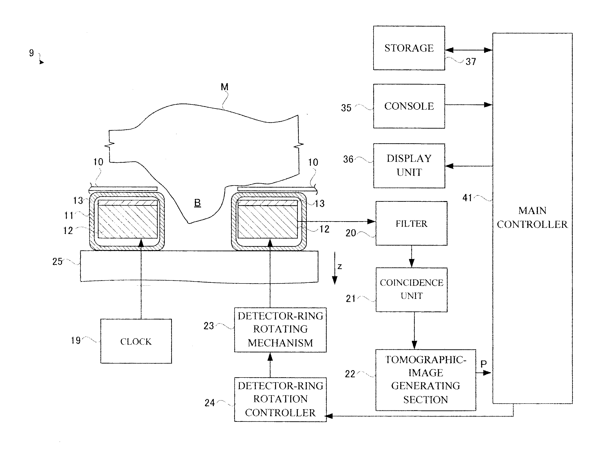
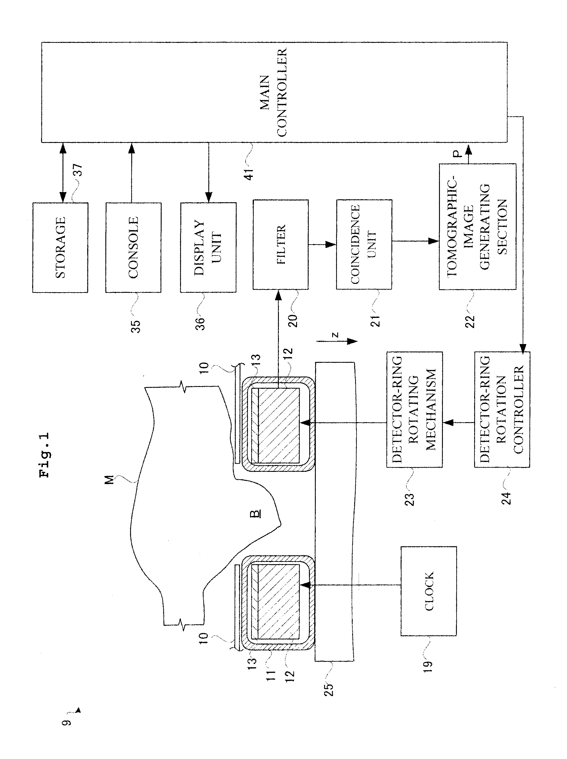
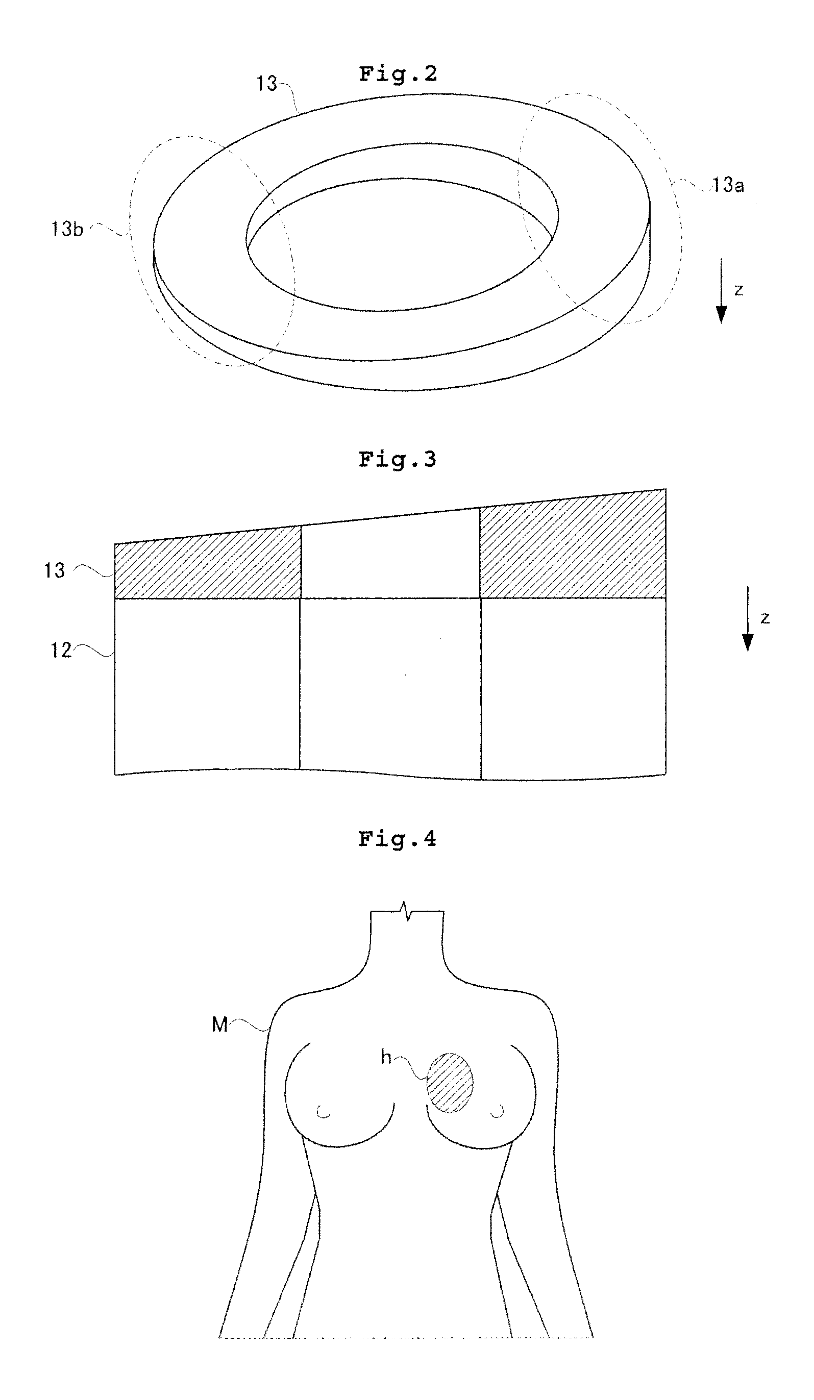
[0036]Examples of a radiation tomographic apparatus according to this invention will be described hereinafter with reference to the drawings. A gamma-ray in Example 1 is one example of radiation in this invention. Here, Example 1 has a construction of a mammography apparatus for breast examination. That is, the radiographic apparatus in Example 1 images radiopharmaceutical distributed in a...


PUM


Abstract

A radiographic apparatus for breast examination, comprising: a detector ring having detectors for detecting radiation being arranged in an arc shape; and a shield plate for absorbing radiation that is provided as to cover a side face of the detector ring perpendicular to a central axis of the detector ring, the shield plate having a thick portion that is thick in a central axis direction and a thin portion that is thin in the central axis direction.

Description

BACKGROUND OF THE INVENTION[0001]1. Field of the Invention[0002]This invention relates to a radiographic apparatus for breast examination that detects annihilation radiation pairs emitted from a subject and images radiopharmaceutical distribution in the subject. Particularly, this invention relates to a radiographic apparatus for breast examination that is used for cancer screening.[0003]2. Description of the Related Art[0004]Medical institutions are equipped with a radiographic apparatus that allows imaging of radiopharmaceutical distribution. Description will be given of a specific configuration of such radiographic apparatus. The conventional radiographic apparatus includes a radiation ring having radiation detectors arranged circularly for detecting radiation. The detector ring detects a pair of radiation (an annihilation radiation-pair), the pair of radiation with opposite directions to each other being emitted from inside of a subject. See, for example, Japanese Patent No. JP-...

Claims


Application Information

Patent Timeline
Patent Type & Authority Applications(United States)
IPC IPC(8): A61B6/04
CPCA61B6/037A61B6/502A61B6/107A61B6/0435
Inventor TONAMI, HIROMICHI
Owner SHIMADZU CORP
Why Eureka
Try Eureka