
Medical device and method for displaying medical image using the same

Inactive Publication Date: 2013-05-16
SAMSUNG ELECTRONICS CO LTD
View PDF8 Cites 6 Cited by
  • Summary
  • Abstract
  • Description
  • Claims
  • Application Information

AI Technical Summary

Benefits of technology

The patent describes a medical device that can display a medical image with an indication of a pathological entity, such as a lesion, on a terminal. The device can receive image information from an external device and display the image on a touch screen. The image can also be enlarged for better visibility when a command is received. The device can also include an input device, such as a remote controller or a voice recognition device, to receive the command. The medical device can also display thumbnails or shortcuts of the image. The patent aims to provide a more efficient way for medical workers to view images that include lesions without having to interpret them redundantly.

Problems solved by technology

Accordingly, although medical imaging devices, such as computer tomography (CT), magnetic resonance imaging (MRI) and X-ray devices have become essential in modern medicine, medical services are often accomplished as a result of an inefficient process requiring a long time and great labor, in which abnormal sites of patients are imaged with medical imaging devices, the images are printed onto films, the films are transferred to an attending physician of the patients, and the attending physician finally performs clinical diagnosis.
As a consequence of this inefficiency, physicians have a reduced capacity for providing rapid and accurate treatment.
In conventional technologies, expensive gateways are required in order to communicate data, or communication is impossible, because consistent standards are not necessarily used and methods for storing and communicating data may vary according to manufacturing companies, or based on the type and the model of imaging devices being used.
Three-dimensional medical images may improve accuracy of lesion detection, but also may entail a longer time for interpretation of medical images and for detection of lesions, due to an increased amount of data which is provided in such images.
The other doctors may check diagnosis results stored by the first doctor, but may inefficiently be required to interpret the three-dimensional medical images again in order to check the corresponding lesions from images.

Method used


Image

  • Medical device and method for displaying medical image using the same
  • Medical device and method for displaying medical image using the same
  • Medical device and method for displaying medical image using the same

Examples


Embodiment Construction

[0055]Hereinafter, exemplary embodiments will be described with reference to the accompanying drawings.

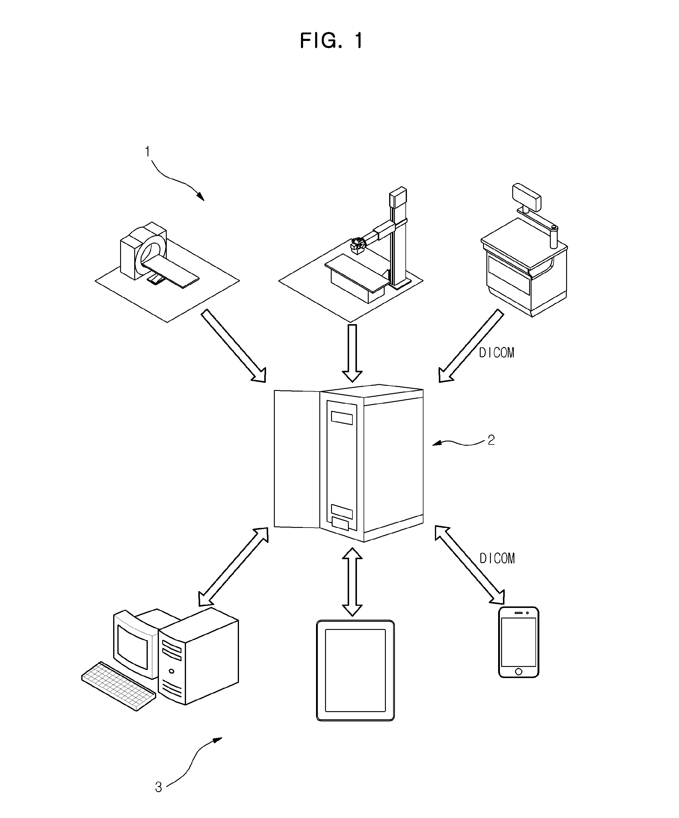
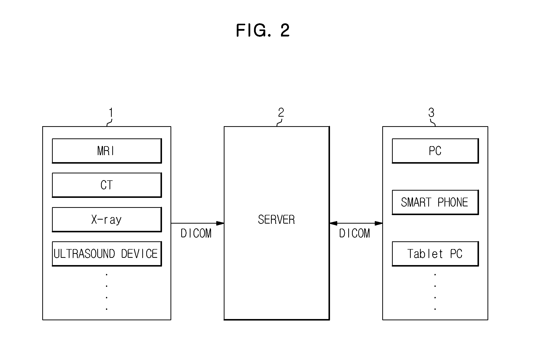
[0056]FIGS. 1 and 2 are views which illustrate a configuration of a medical imaging system according to an exemplary embodiment.

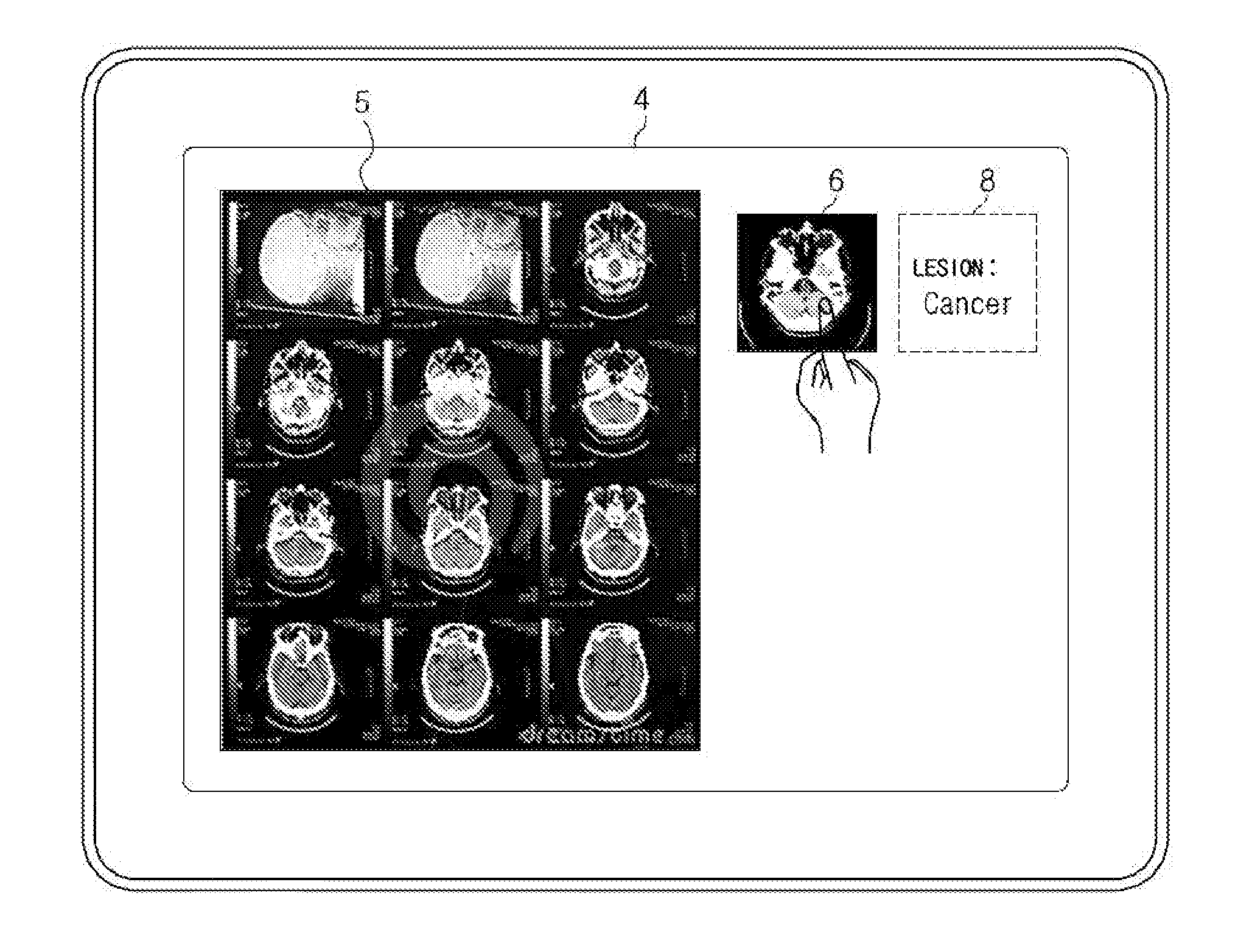
[0057]As shown in FIG. 1, the medical imaging system includes an image acquisition device 1 which acquires an image relating to a subject, a server 2 which receives the acquired image from the image acquisition device 1 and stores the received image in a medical display device 3, and the medical display device 3 which receives the image relating to the subject from the server 2 and displays the image.

[0058]As shown in FIG. 2, the image acquisition device 1 acquires three-dimensional images relating to subjects to be imaged, and may include, for example, a MRI device, a CT device, an X-ray device, and / or an ultrasound device or the like.

[0059]The image acquisition device 1 transfers three-dimensional images relating to the subject which are acquired by exec...


PUM


Abstract

Disclosed is a medical device which displays an object relating to an image considered to include an indication relating to a pathological entity, such as, for example, a lesion, on a terminal on which a medical image is displayed, and a method for displaying a medical image by using the same. The medical device includes a display device, a communication device which communicates with an external device, and a controller which displays an image which includes the indication relating to a pathological entity on the display device when the communication device receives image information relating to a subject.

Description

CROSS-REFERENCE TO RELATED APPLICATION[0001]This application claims priority from Korean Patent Application No. 10-2011-0118911, filed on Nov. 15, 2011, in the Korean Intellectual Property Office, the disclosure of which is incorporated herein by reference in its entirety.BACKGROUND[0002]1. Field[0003]Exemplary embodiments relate to a medical device. In particular, exemplary embodiments relate to a medical device and a method for displaying a medical image by using the same, in order to reduce medical image interpretation time required by medical workers and thereby provide efficient diagnosis.[0004]2. Description of the Related Art[0005]In general, clinical diagnosis has an important role in treatment of patients in medical operations, development of medical techniques greatly contributes to accurate clinical diagnosis, and dependence thereon will continue to increase.[0006]Accordingly, although medical imaging devices, such as computer tomography (CT), magnetic resonance imaging (...

Claims


Application Information

Patent Timeline
IPC IPC(8): A61B6/00G16H10/60
CPCA61B6/463A61B5/0013A61B6/466A61B6/467A61B6/5217A61B6/563A61B6/566A61B5/0033A61B8/463A61B8/465A61B8/466A61B8/467A61B8/565A61B6/548A61B6/465G06F3/14H04B7/24
Inventor SHIN, SEUNG WOO
Owner SAMSUNG ELECTRONICS CO LTD
Features
  • Generate Ideas
  • Intellectual Property
  • Life Sciences
  • Materials
  • Tech Scout
  • Unparalleled Data Quality
  • Higher Quality Content
  • 60% Fewer Hallucinations
Learn More