System and Method for Denoising Medical Images Adaptive to Local Noise

Active Publication Date: 2013-08-08
MAYO FOUND FOR MEDICAL EDUCATION & RES
View PDF1 Cites 24 Cited by
  • Summary
  • Abstract
  • Description
  • Claims
  • Application Information

AI Technical Summary

Benefits of technology

[0013]The present invention overcomes the aforementioned drawbacks by providing a system and method that can efficiently estimate the local noise of CT images,

Problems solved by technology
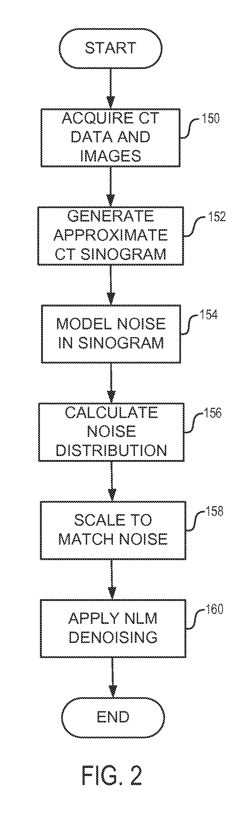
The drastically increased use of CT in modern clinical settings has generated serious public health concerns regarding the cancer risks associated with the radiation exposure from CT.
However, lowering radiation dose alone generally produces a noisier image and may seriously degrade diagnostic performance.
One potential drawback of projection-based methods is that they may result in some loss of image sharpness due to the fact that edges in projection data are not well-defined.
However, CT noise in image space is difficult to model accurately and has strong spatial variations and correlations.
It can therefore be more difficult for such techniques to achieve an optimal tradeoff between denoising and blurring or artifacts, or to get consistent performance across an entire scan volume.
True IR is very computationally intensive (e.g., several hours per data set), w

Method used


Image

  • System and Method for Denoising Medical Images Adaptive to Local Noise
  • System and Method for Denoising Medical Images Adaptive to Local Noise
  • System and Method for Denoising Medical Images Adaptive to Local Noise

Examples


Embodiment Construction

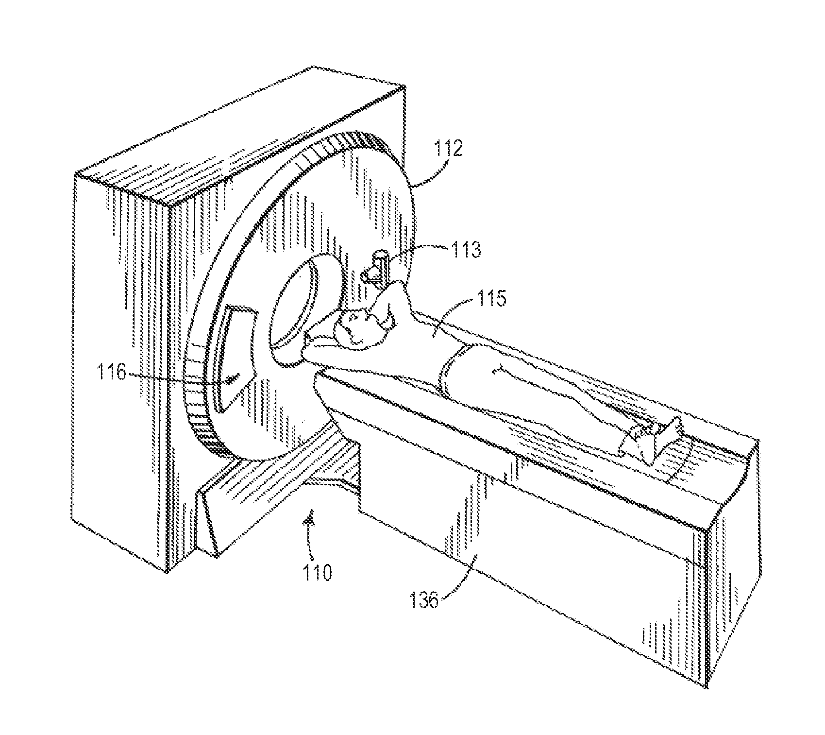
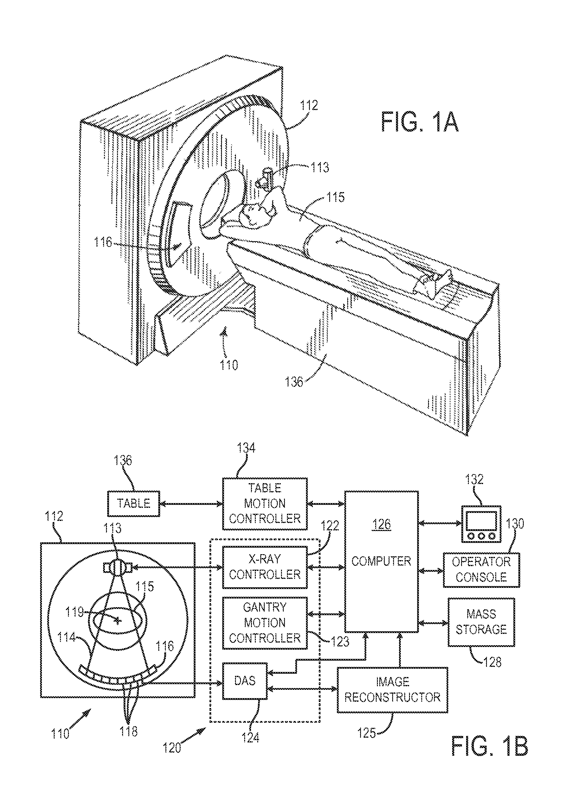
[0020]With initial reference to FIGS. 1A and 1B, a computed tomography (CT) imaging system 110 includes a gantry 112 representative of at least a “third generation” CT scanner. In the illustrated example, the gantry 112 has a pair of x-ray sources 113 that each project a fan beam or cone beam of x-rays 114 toward a detector array 116 on the opposite side of the gantry 112. The detector array 116 is formed by a number of detector elements 118 that together sense the projected x-rays that pass through a medical patient 115. During a scan to acquire x-ray projection data, the gantry 112 and the components mounted thereon rotate about a center of rotation 119 located within the patient 115 to acquire attenuation data.

[0021]The rotation of the gantry 112 and the operation of the x-ray source 113 are governed by a control mechanism 120 of the CT system 110. The control mechanism 120 includes an x-ray controller 122 that provides power and timing signals to the x-ray sources 113 and a gant...


PUM


Abstract

A system and method is provided for estimating the local noise of CT images and denoising the images using a modified non-local means (NLM) algorithm that is adaptive to local variations of noise levels. A strategy for efficiently estimating the local noise of CT images is also described.

Description

CROSS-REFERENCE TO RELATED APPLICATIONS[0001]The present application is based on, claims priority to, and incorporates herein by reference in its entirety U.S. Provisional Patent Application No. 61 / 595,999, filed Feb. 7, 2012, and entitled “SYSTEM AND METHOD FOR CONTROLLING RADIATION DOSE FOR RADIOLOGICAL APPLICATIONS.”BACKGROUND OF THE INVENTION[0002]The present invention relates to medical imaging and, more particularly, to systems and methods for controlling radiation doses delivered when performing imaging processes using ionizing radiation.[0003]In a computed tomography system, an x-ray source projects a beam which is collimated to lie within an X-Y plane of a Cartesian coordinate system, termed the “imaging plane.” The x-ray beam passes through the object being imaged, such as a medical patient or other non-medical patient or object, such as in industrial CT imaging, and impinges upon an array of radiation detectors. The intensity of the transmitted radiation is dependent upon...

Claims


Application Information

Patent Timeline
IPC IPC(8): A61B6/00A61B6/03
CPCA61B6/5258G06T11/003A61B6/583A61B6/5205A61B6/542A61B6/032
Inventor YU, LIFENGMANDUCA, ARMANDOLI, ZHOUBOFLETCHER, JOEL G.MCCOLLOUGH, CYNTHIA H.
Owner MAYO FOUND FOR MEDICAL EDUCATION & RES
Why Eureka
Try Eureka