

Microfluidic device for generating neural cells to simulate post-stroke conditions
a microfluidic device and post-stroke technology, applied in the direction of fluorescence/phosphorescence, apoptosis detection, instruments, etc., can solve the problem of extremely limited treatment options for ischemic strok
- Summary
- Abstract
- Description
- Claims
- Application Information
AI Technical Summary
Benefits of technology
Problems solved by technology
Method used
Image

Examples
examples
I. Fabrication of a Microfluidic Cell Culture Device
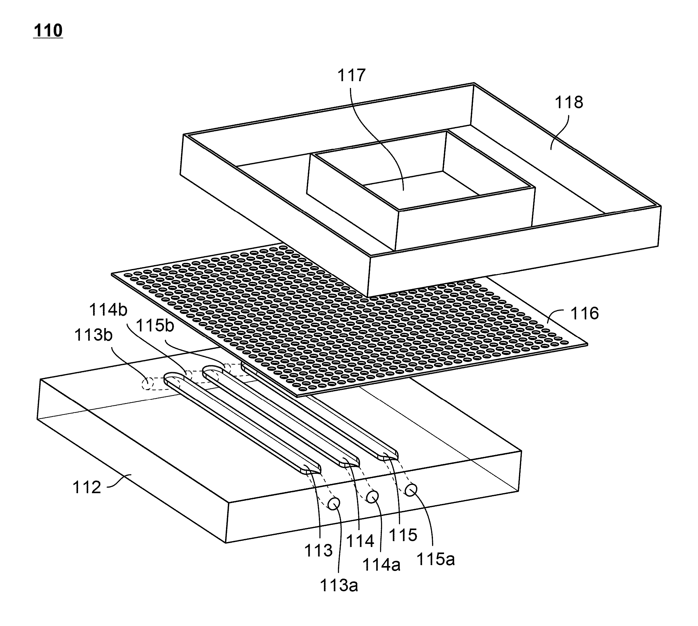
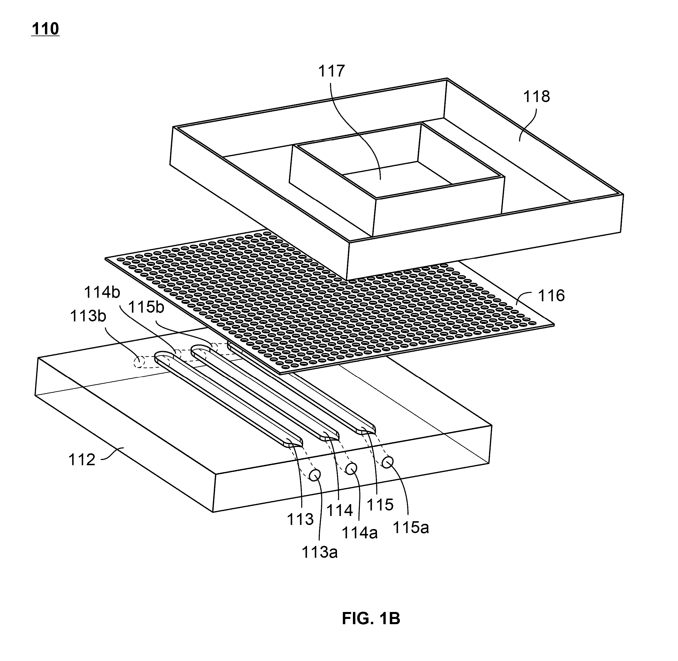
[0109]A microfluidic device for subjecting different neural cells to different environments can be produced according to the methods below. This device bears a neural compartment juxtaposed above three parallel microchannels. The neural compartment is separated from the microchannels by a microporous polyester membrane bonded irreversibly through a thin layer of silicone adhesive. FIG. 1 shows an exploded view of such a device.
[0110]The neural chamber accommodates a cell culture area of ˜0.5 cm2, close to the surface area of a section of adult rat brain. The channels are fabricated to have dimensions of 1 mm width, 250 μm height and 1.1 cm (length) separated from each other by 1 mm. The design of the neural chamber facilitates standard well-plate format of cell culture, which is simple, routine, and does not require continuous perfusion. The reservoir in the culture chamber holds ˜150 μl media. The membrane (10 μm thickness and por...
PUM
| Property | Measurement | Unit |
|---|---|---|
| diameter | aaaaa | aaaaa |
| diameter | aaaaa | aaaaa |
| temperature | aaaaa | aaaaa |
Abstract
Description
Claims
Application Information