Minimally invasive neurosurgical intracranial robot system and method

a robot system and neurosurgical technology, applied in the field of minimally invasive neurosurgical procedures, can solve the problems of high mortality rate, lack of satisfactory continuous imaging modality for intraoperative intracranial procedures, and extremely difficult to remove brain tumors precisely and completely without damaging the surrounding brain tissue using traditional surgical tools, and achieve high efficiency and precise manner.

Inactive Publication Date: 2013-08-22
UNIV OF MARYLAND
View PDF12 Cites 58 Cited by
  • Summary
  • Abstract
  • Description
  • Claims
  • Application Information

AI Technical Summary

Benefits of technology

[0030]It is therefore an object of the present invention to provide a robotic system for minimally invasive surgical procedures which can be teleoperatively controlled by a neurosurgeon in a highly efficient and precise manner.

Problems solved by technology

The primary reason for the high mortality rate includes the inability to remove the complete tumor tissue due to the location of the tumor deep in the brain, as well as the lack of a satisfactory continuous imaging modality for intraoperative intracranial procedures.
However, due to the lack of satisfactory continuous imaging modality, it is extremely challenging to remove brain tumors precisely and completely without damaging the surrounding brain tissue using traditional surgical tools.
As a result, patients may develop hemi paresis, cognitive impairment, stroke or other neurological deficits due to the procedure.
However, due to the strong magnetic field required in the MRI, commonly used sensors and actuators in conventional robotic systems are precluded from being used in MRI-compatible robots.
Although the above-mentioned robotic systems are MRI compatible, they unfortunately cannot be used to reach a target which is not in the “line-of-sight” due to limited Degrees Of Freedom (DOF) of the robots intended for use in their systems.
Though the approach of using SMA (Shape Memory Alloy) wires as actuators was successful, there are significant limitations.
The current can interfere with the magnetic field inside the MRI bore, and thus may lead to some distortion in the image.
1153-1158), the noise and distortion might still cause difficulties finding precise tumor boundaries.

Method used


Image

  • Minimally invasive neurosurgical intracranial robot system and method
  • Minimally invasive neurosurgical intracranial robot system and method
  • Minimally invasive neurosurgical intracranial robot system and method

Examples


Embodiment Construction

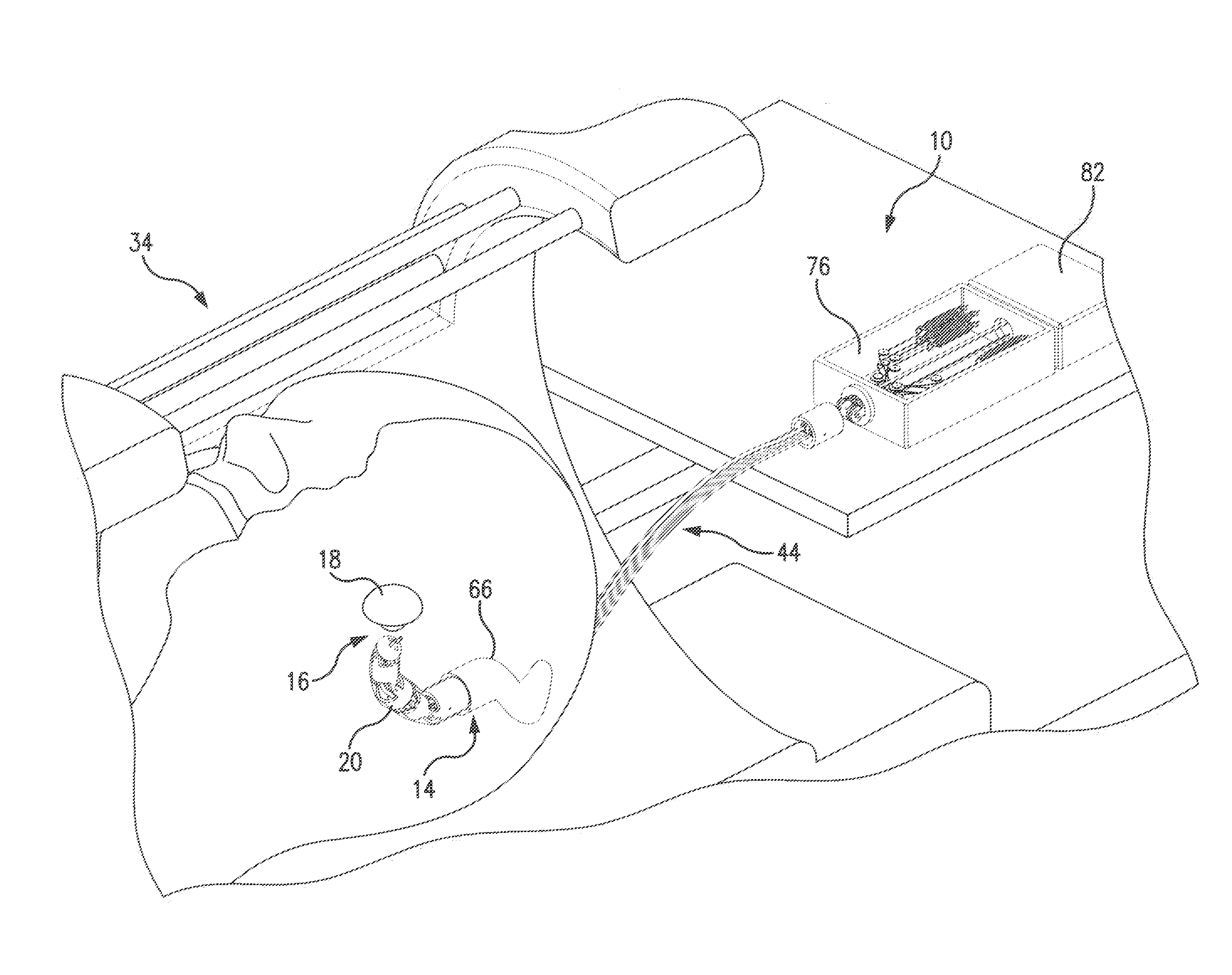
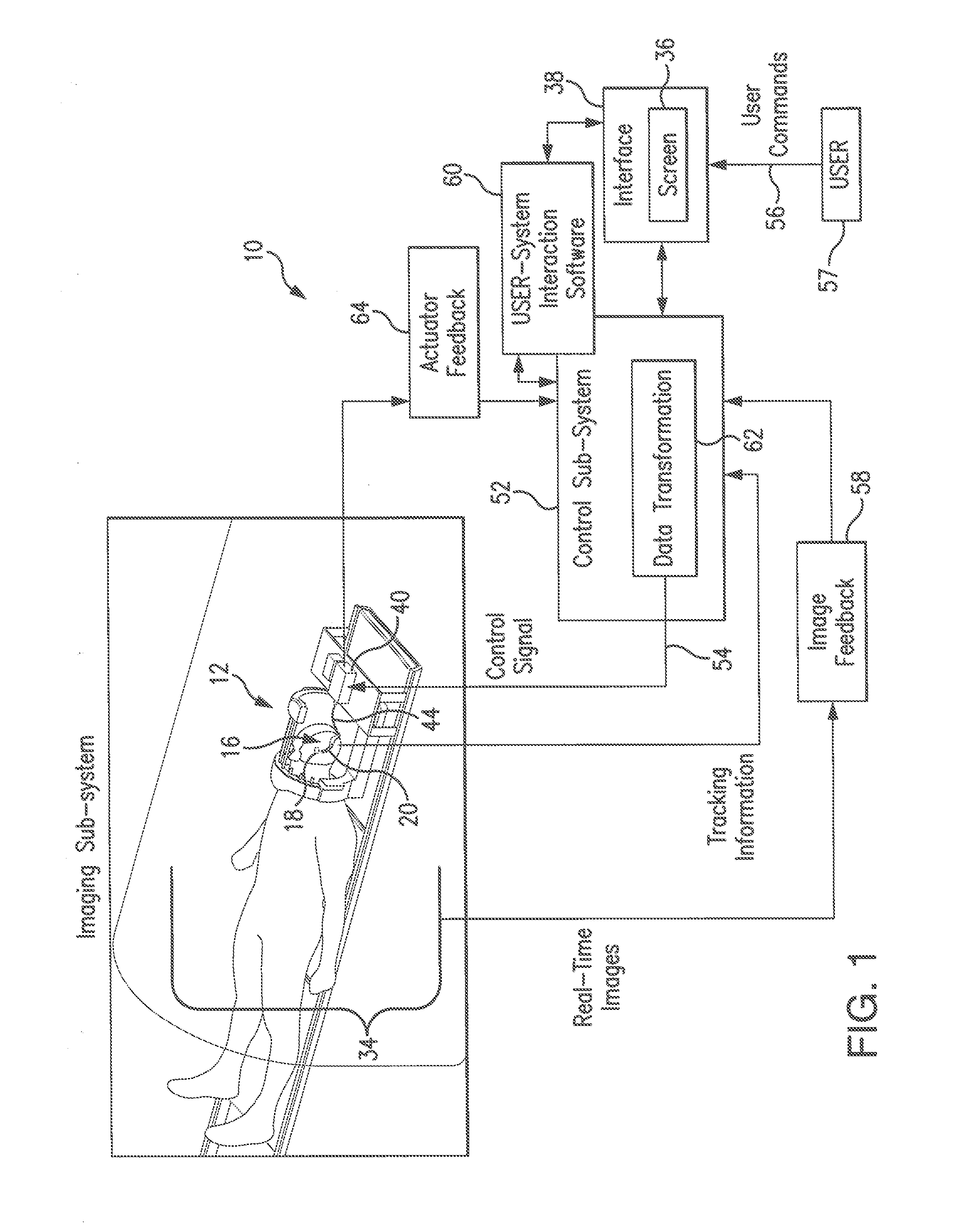
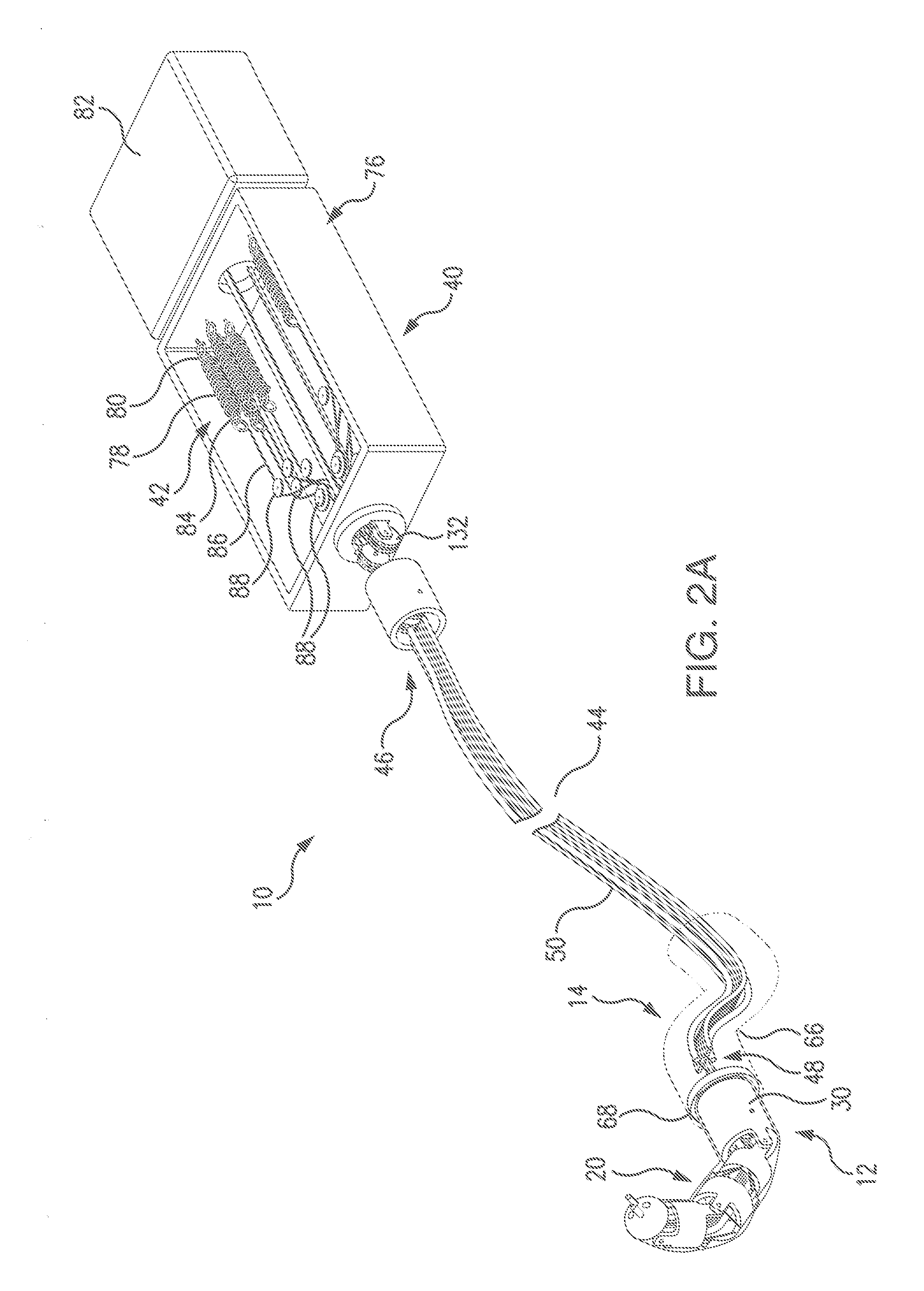
[0116]Referring to FIG. 1, a minimally invasive neurosurgical intracranial robot (also referred to herein as MINIR) system 10 includes a robot sub-system 12, which during the surgery is introduced through a narrow surgical corridor to an operative site 16 containing a tumor 18.

[0117]The present system will enable the neurosurgeon to remove deep brain intracranial tumors, both neoplastic and non-neoplastic masses, such as blood clots, that are typically hard to reach through a minimally invasive approach, since deep brain tumors are typically located away from the “line-of-sight” of the neurosurgeon. The present system provides the neurosurgeon with means to remove the tumor by teleoperatively navigating the position of the minimally invasive neurosurgical intracranial robot (MINIR) having a number of DOFs (Degrees-of-Freedom) towards the tumor site based on real-time images of the operational site aligned with visual tracking data of the robot, as will be presented in detail in furt...


PUM


Abstract

Minimally invasive neurosurgical intracranial robot system is introduced to the operative site by a neurosurgeon through a narrow surgical corridor. The robot is passed through a cannula and is attached to the cannula by a latching mechanism. The robot has several links interconnected via revolute joints which are tendon-driven by tendons routed through channels formed in the walls of the links. The robot is teleoperatively guided by the neurosurgeon based on real-time images of the intracranial operative site and tracking information of the robot position. The robot body is equipped with a tracking system, tissue liquefacting end-effector, at as well as irrigation and suction tubes. Actuators for the tendon-driven mechanism are positioned at a distance from the imaging system to minimize distortion to the images. The tendon-actuated navigation of the robot permits an independent control of the revolute joints in the robot body.

Description

REFERENCE TO RELATED APPLICATIONS[0001]This Utility patent application is based on the Provisional Patent Application No. 61 / 596,603 filed on 8 Feb. 2012.STATEMENT REGARDING FEDERAL RESPONSIVE RESEARCH OR DEVELOPMENT[0002]The development of the invention described herein was funded by NIH under Grant No. R21EB008796. The U.S. Government has certain rights in this invention.FIELD OF THE INVENTION[0003]The present invention relates to minimally invasive surgical procedures, and more in particular to a robotic system for minimally invasive neurosurgery.[0004]Even more in particular, the present invention relates to a high dexterity robot for applications in neurosurgery, which can be teleoperatively controlled by a neurosurgeon to remove deep intracranial tumors, both neoplastic and non-neoplastic masses, such as blood clots.[0005]The present invention further relates to a miniature robot teleoperatively guided by a neurosurgeon based on the images of the intracranial operation site ac...

Claims


Application Information

Patent Timeline
Patent Type & Authority Applications(United States)
IPC IPC(8): A61B19/00A61M1/00A61B18/12A61B18/04A61B8/08A61N7/00A61B5/055A61B5/06A61B6/03A61B17/00A61B18/20
CPCA61B19/5244A61B6/12A61N7/022A61B18/1492A61B2017/00477A61B2018/00446A61B2019/2238A61B2019/2246A61B19/2203A61B17/00234A61M1/0058A61B18/12A61B18/042A61B18/20A61N7/00A61B5/055A61B5/062A61B6/032A61B8/0841A61B2018/00577A61B2019/5251A61B2034/715A61B34/20A61B34/30A61B2034/306A61B2034/2051A61M1/77
Inventor DESAI, JAYDEV P.HO, MINGYENSIMARD, J. MARCGULLAPALLI, RAO
Owner UNIV OF MARYLAND
Why Eureka
Try Eureka