Spherical NANO and microparticles derived from plant viruses for the display of foreign proteins or epitopes

a technology of plant viruses and nanoparticles, which is applied in the direction of viral antigen ingredients, peptide sources, carrier-bound antigen/hapten ingredients, etc., can solve the problems of genetic instability, labor- and time-consuming, and the study was not developed later

Inactive Publication Date: 2013-09-12
ATABEKOV IOSIF GRIGORIEVICH +7
View PDF0 Cites 0 Cited by
  • Summary
  • Abstract
  • Description
  • Claims
  • Application Information

AI Technical Summary

Benefits of technology

[0084]On the whole, fast, simple and widely available methods for (i) new stable, universal and biologically safe virus-generated SPs platform production and for (ii) in vitro assembly of SPs-based immunogenic compositions with adjuvant activity were developed.

Problems solved by technology

Unfortunately, these studies were not developed later on.
1. Construction of chimeric viruses by the methods of genetic engineering is labour- and time-consuming (about 3-10 months) for modification the viral genome and creation the recombinant genome producing chimeric CP fused genetically to a foreign antigen / epitope.
2. Not infrequently chimeric viruses cannot systemically infect the plants, but are genetically unstable, i.e. they revert into the wild type or loose the foreign epitopes in the course of replication and cell-to-cell movement.
The disadvantage of affinity-conjugated antigen system is the complexity of the procedure of making two components: e.g. the biotin-decorated recombinant virus and the “SA-antigen” complex exhibiting a specific affinity to each other.
The limitation of affinity-conjugated antigen system is due to the fact that specific affinity exists only between two moieties: the modified platform (viral CP) and SA-antigen.

Method used


Image

  • Spherical NANO and microparticles derived from plant viruses for the display of foreign proteins or epitopes
  • Spherical NANO and microparticles derived from plant viruses for the display of foreign proteins or epitopes
  • Spherical NANO and microparticles derived from plant viruses for the display of foreign proteins or epitopes

Examples


example 1

Native Tobamoviruses and Different Forms of TMV CP Aggregates

[0145]TMV U1 strain was isolated from systemically infected Nicotiana tabacum L. cv. Samsun plants as described previously (Novikov, V. K. and Atabekov, J. G. (1970) A study of the mechanism controlling the host range of plant virus. I Virus—specific receptors of Chenopodium amaranticolor. Virology 41, 101-107).

[0146]Cucumber green mottle mosaic virus (CGMMV) was isolated from the extracts of systemically infected cucumber plants, clarified at 10,000 g, emulgated with chlorophorm (30 min) and centrifuged at 400 rpm for 30 min. Then polyethileneglicol (PEG 6000) was added (3%) to supernatant with 0.2M NaCl and incubated for night at 40%. After centrifugation at 10,000 g the pellet was resuspended in 0.02 M Tris-HCl, 0.001M EDTA, pH 7.5 and clarified at 10,000 g. The extraction from the pellet was repeated twice. The supernatants were mixed and subjected to two cycles of differential centrifugation (for 2 h at 45,000 rpm in ...

example 2

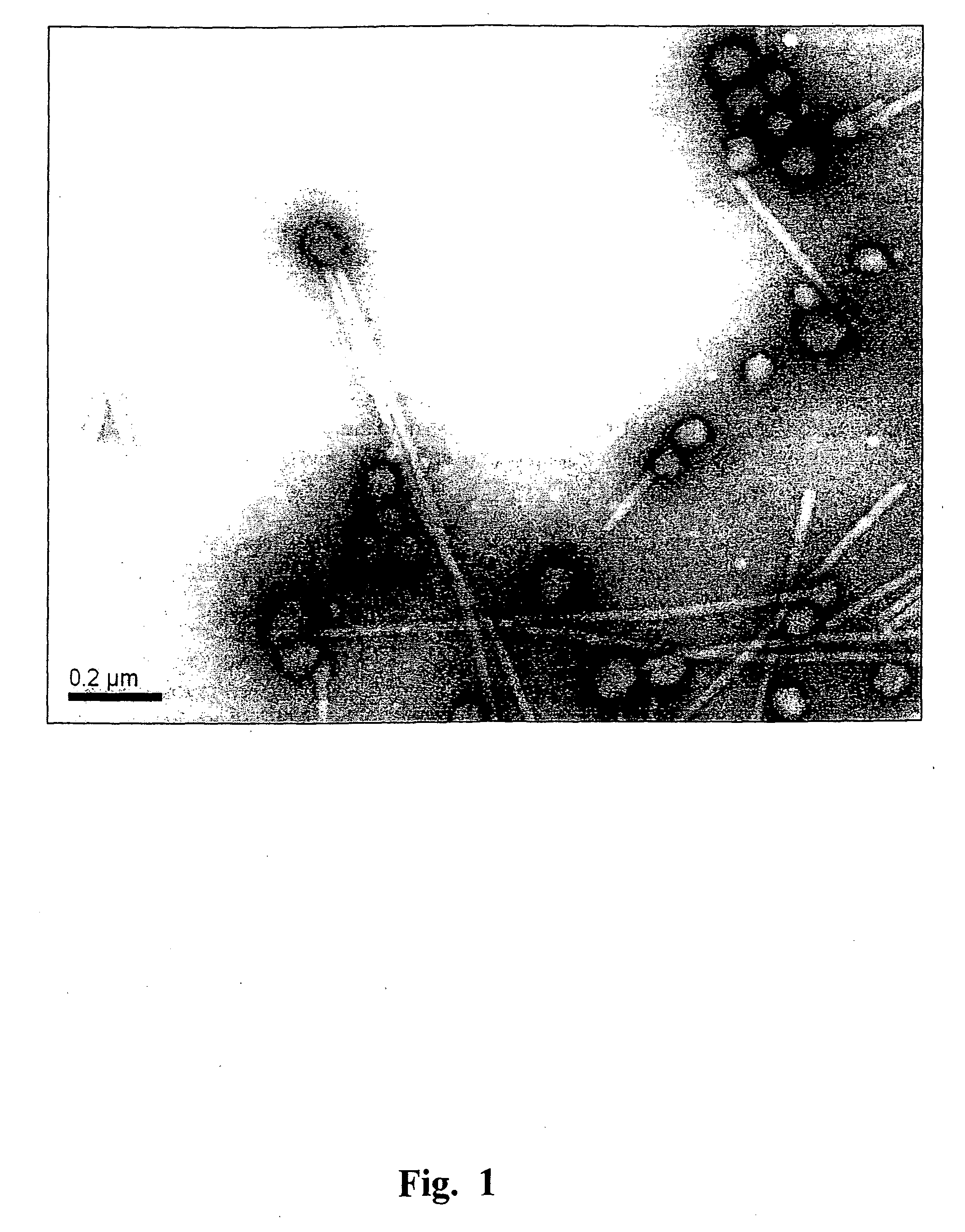
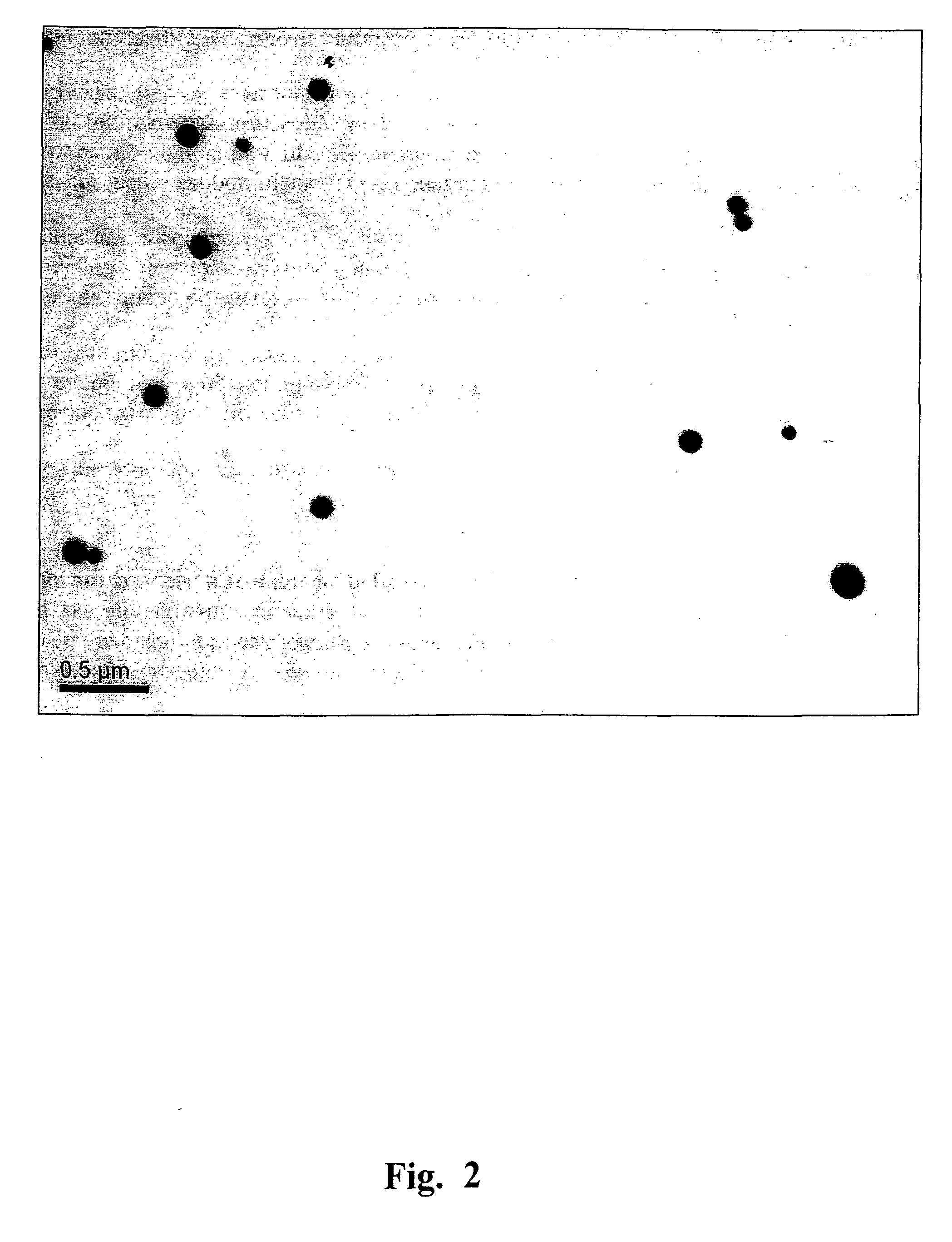
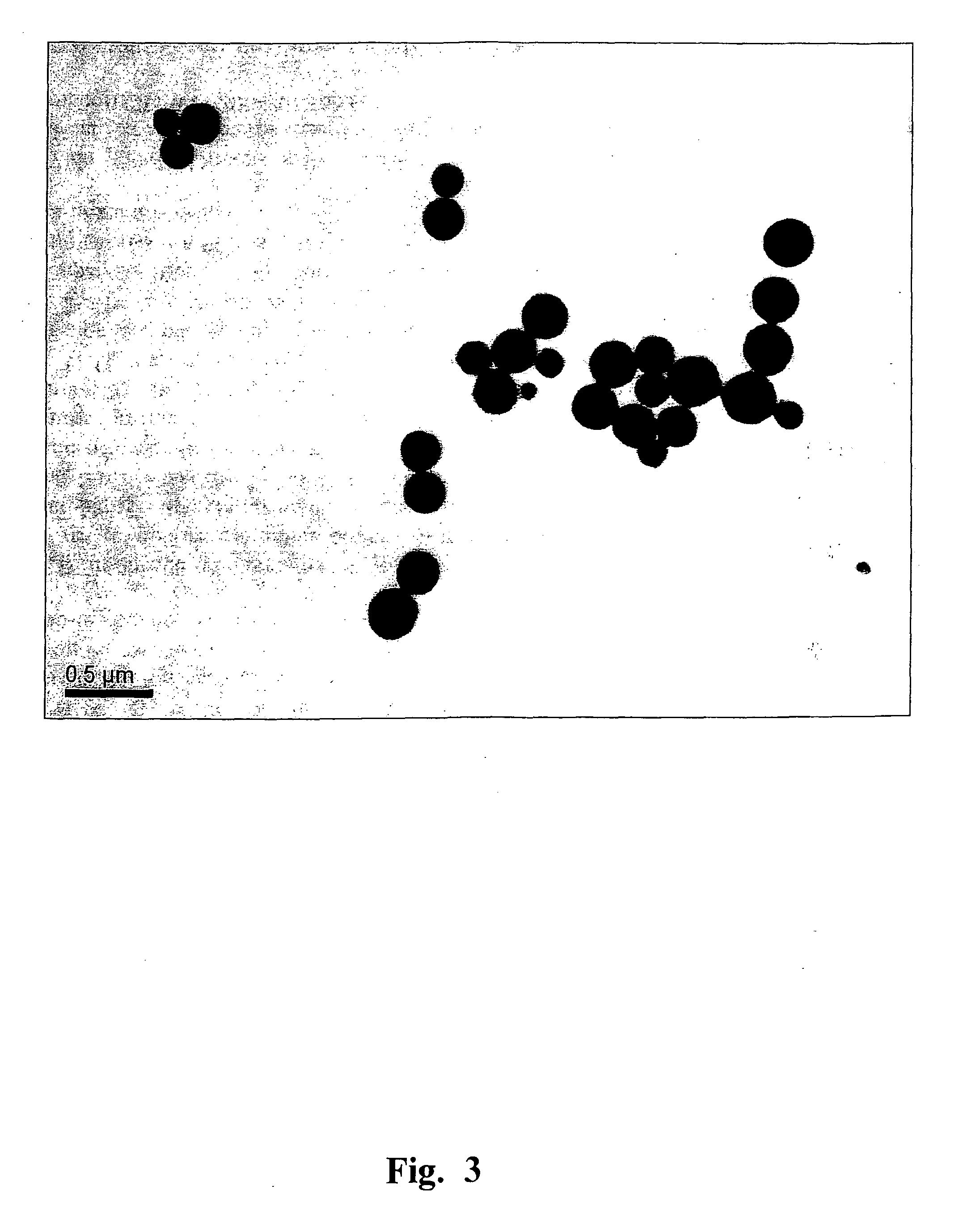
Generation of SPs and their Examination by Transmission and Scanning Electron Microcopy

[0148]Heating of TMV and viral RNA-free CP preparations was performed in the “Tercyc” thermocycler (“DNA-technology”, Russia) for 10 sec at the temperature required. The process of the native TMV into SP transition was examined by TEM and SEM. The specimens were prepared as reported previously (Kaftanova A. S., Kiselev N. A., Novikov V. K., Atabekov J. G. (1975) Structure of products of protein reassembly and reconstitution of potato virus X. Virology 65, 283-287). The samples were viewed using a JEOL JEM-1011 microscope (JEOL, Japan) operating at 80 kV. Digital images were captured using a Gatan Erlangshen ES500W camera and Gatan Digital Micrograph™ software. For SEM, a drop of the sample was placed on a specimen stub and dried in air, then sputtered with gold with palladium in an ionic sputtering installation IB-3 Ion Coater (Eico, Japan), and examined in microscope JSM-6380LA (JEOL, Japan). FIG...

example 3

SPs Generation by RNA-Free Preparations of TMV CP

[0149]FIG. 8 presents a schematic (not to scale) representation of the SPs generation by native TMV and by RNA-free forms of TMV protein. The numbers indicate concentrations of native TMV (at left) and RNA-free TMV proteins (at right) heated at 94° C. or 65° C., respectively. The size ranges of SP (in nm) are indicated.


PUM

PropertyMeasurementUnit
temperatureaaaaaaaaaa
sizeaaaaaaaaaa
sizeaaaaaaaaaa

Abstract

A novel type of particle platform for application in biotechnology is provided by the present invention. Said platforms comprise RNA-free particles generated by thermal denaturation and structural remodeling of helical plant viruses. Tobamovir-uses and, in particular, tobacco mosaic virus (TMV) coat protein (CP) subunits, denatured, at high temperatures are specifically self-assembled by two-stage assembly into the spherical particles (SPs) of similar shape and varying size including nanoparticles (SNPs) with a diameter up to 100-150 nm and spherical microparticles (SMPs) with a diameter up to 800 nm and more. The size of said SPs depends on the virus concentration used and, therefore can be controlled. Said SPs are biologically safe, highly stable and highly immunogenic. Said SNPs and SMPs are structurally distinct from viruses presently known. They are unique, having no protein nano-particle analogs in the nature. Said particles can be produced by the native virus and also by different forms of viral CP lacking RNA. The SPs can be generated by thermal denaturation and structural remodeling of different helical plant viruses belonging to genera Tobamovirus, Hordeivirus and family Flexiviridae. The invention relates to the creation of functionally active compostions on the base of said SP-platforms for use in medicine, veterinary, virology, immunology and diagnostics.
In accordance with this aspect of the present invention, several immunogenic compositions were obtained comprising SP platform and linked to the SP surface foreign full-size proteins, including green fluorescent protein, coat protein of PVX or epitopes of several different viruses (e.g. human influenza A virus and rubella virus epitopes). The procedure of in vitro assembly of said type of composition took about one hour. Apparently, the nanocomplexes of SNP/SMP with foreign antigens/epitopes could be regarded as candidate nanovaccines. In accordance with another aspect of the present invention, we found that the TMV-generated SPs can be used as immunological booster or adjuvant stimulating the immune response of animals by parenteral immunization.

Description

TECHNICAL FIELD[0001]The present invention relates to the field of nanocomplexes and microcomplexes production by introducing in biotechnology a novel type of particle platform for assembly of biologically active compositions, and using said compositions in medicine, veterinary, virology, immunology and diagnostics.BACKGROUND ART[0002]Said particle platforms comprise spherical particles (SPs) of different size including nanoparticles (SNPs) or microparticles (SMPs) obtained by thermal denaturation and structural remodeling of a native helical plant virus particles or RNA-free coat protein (CP) isolated from the native virus. More precisely, said SPs comprise thermally denatured and specifically assembled into spherical particles viral coat protein subunits. SPs are completely biologically safe, highly immunogenic, stable, and inexpensive. The size of SPs depends on virus concentration and, therefore can be controlled.[0003]SNP / SMP are unique and have no structural analogs among viru...

Claims


Application Information

Patent Timeline
Patent Type & Authority Applications(United States)
IPC IPC(8): C07K14/08A61K39/39
CPCA61K2039/5258C12N2770/26062C07K14/005C07K14/08C07K17/00A61K2039/6075A61K2039/64C07K2319/20C07K2319/21C12N2760/16134C12N2770/36234C12N2770/40022C12N2770/40042C12N2770/40062A61K39/39A61K39/145A61K39/12A61K39/385C12N2770/26022C12N2770/26023C12N2770/26042C12N2770/34034B82Y5/00
Inventor ATABEKOV, IOSIF GRIGORIEVICHKARPOVA, OLGA VIACHESLAVOVNAKIRPICHNIKOV, MIKHAIL PETROVICHNIKITIN, NIKOLAY ALEXANDROVICHCHIRKOV, SERGEY NIKOLAEVICHTRIFONOVA, EKATERINA ALEXEEVNASHEVELEVA, ANNA ALEXANDROVNAARCHIPENKO, MARINA VLADIMIROVNA
Owner ATABEKOV IOSIF GRIGORIEVICH
Why Eureka
Try Eureka