Motion Compensated MR Imaging System

a motion correction and imaging system technology, applied in the field of motion correction mr image data, can solve the problems of respiratory related patient physical motion unavoidable during mr imaging, reduce image quality for clinical diagnosis, and inconvenient collection of mr data, so as to reduce respiratory related motion artifacts, reduce respiratory related motion, and reduce the effect of patient effor

Active Publication Date: 2013-09-26
SIEMENS HEALTHCARE GMBH
View PDF4 Cites 31 Cited by
  • Summary
  • Abstract
  • Description
  • Claims
  • Application Information

AI Technical Summary

Benefits of technology

[0007]An MR imaging system reduces respiratory related motion artifacts using measured information while demanding little patient effort and cooperation with minimal impact on image processing time and provides improved image quality in clinical abdominal MR applications, for example. A method provides motion corrected MR image data in an MR imaging system. The method employs an imaging method for acquiring k-space data of a k-space data array during an imaging scan. The k-space data represents image data of a patient anatomical region. The method acquires k-space data associated with a central region of the k-space data array and subsequently acquires k-space data external to the central region of the k-space data array. The method also acquires a motion signal indicating respiratory motion at least during the acquisition of the k-space data external to the central region and compares the motion signal with a predetermined threshold. The method identifies acquired k-space data corresponding to acquisition periods when the motion signal exceeds the threshold and excludes use of the identified acquired k-space data in image reconstruction using the remaining acquired k-space data.

Problems solved by technology

Respiration related MR imaging artifacts occur if they are not suppressed or reduced, which leads to ghosting that may overlay a region of interest or blurring which reduces image quality for clinical diagnosis.
Respiration related patient physical motion is unavoidable during MR imaging, especially for body region imaging, such as of the abdomen, spine, pelvis and heart.
During a respiration cycle, the physical position of many organs, such as of a heart, liver or diaphragm changes resulting in inconsistency in collected MR data (phase shifts) during MR data acquisition.
However, for many patients, a long breathhold may be impossible to achieve, depending on patient physical condition, age and health.
This method is relatively inefficient resulting in prolonged scanning time and increase in patient discomfort.
This method also typically results in variable image contrast due to long and variable repetition time (TR).
The use of segmented acquisition improves data collection efficiency at the cost of image quality reduction by allowing organ motion within a certain range but the scanning time may still be too long for some patients.
Selection and discarding “bad” data during a late phase of breathhold is important in this method and reliability of the known system is largely dependent on the acquired signal quality, i.e. signal-noise-ratio (SNR) and the method is vulnerable to a poor SNR which is common for non-central k-space data.

Method used


Image

  • Motion Compensated MR Imaging System
  • Motion Compensated MR Imaging System
  • Motion Compensated MR Imaging System

Examples


Embodiment Construction

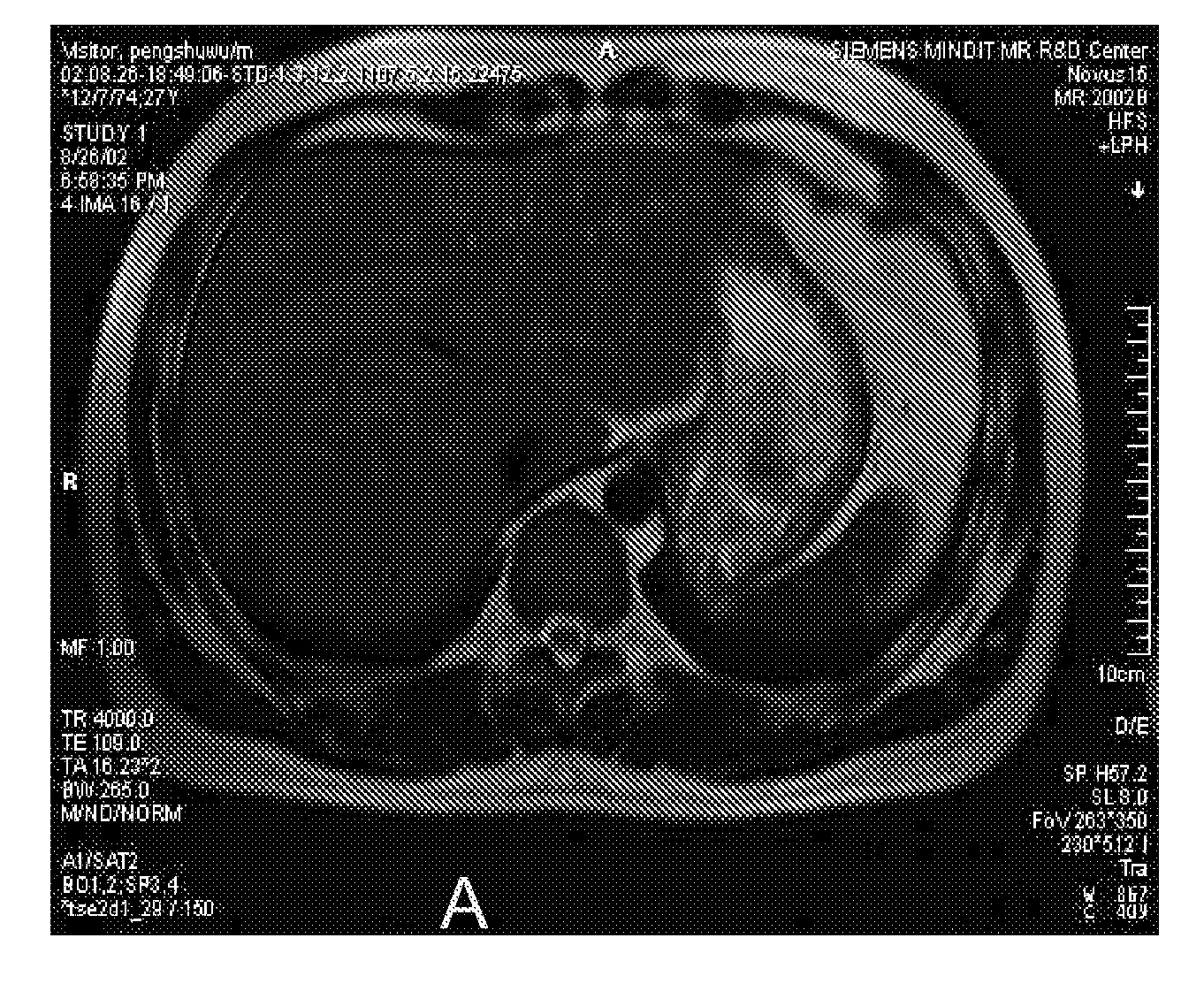
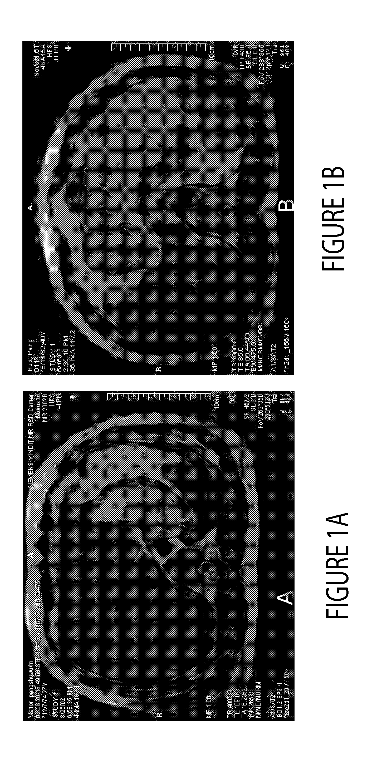
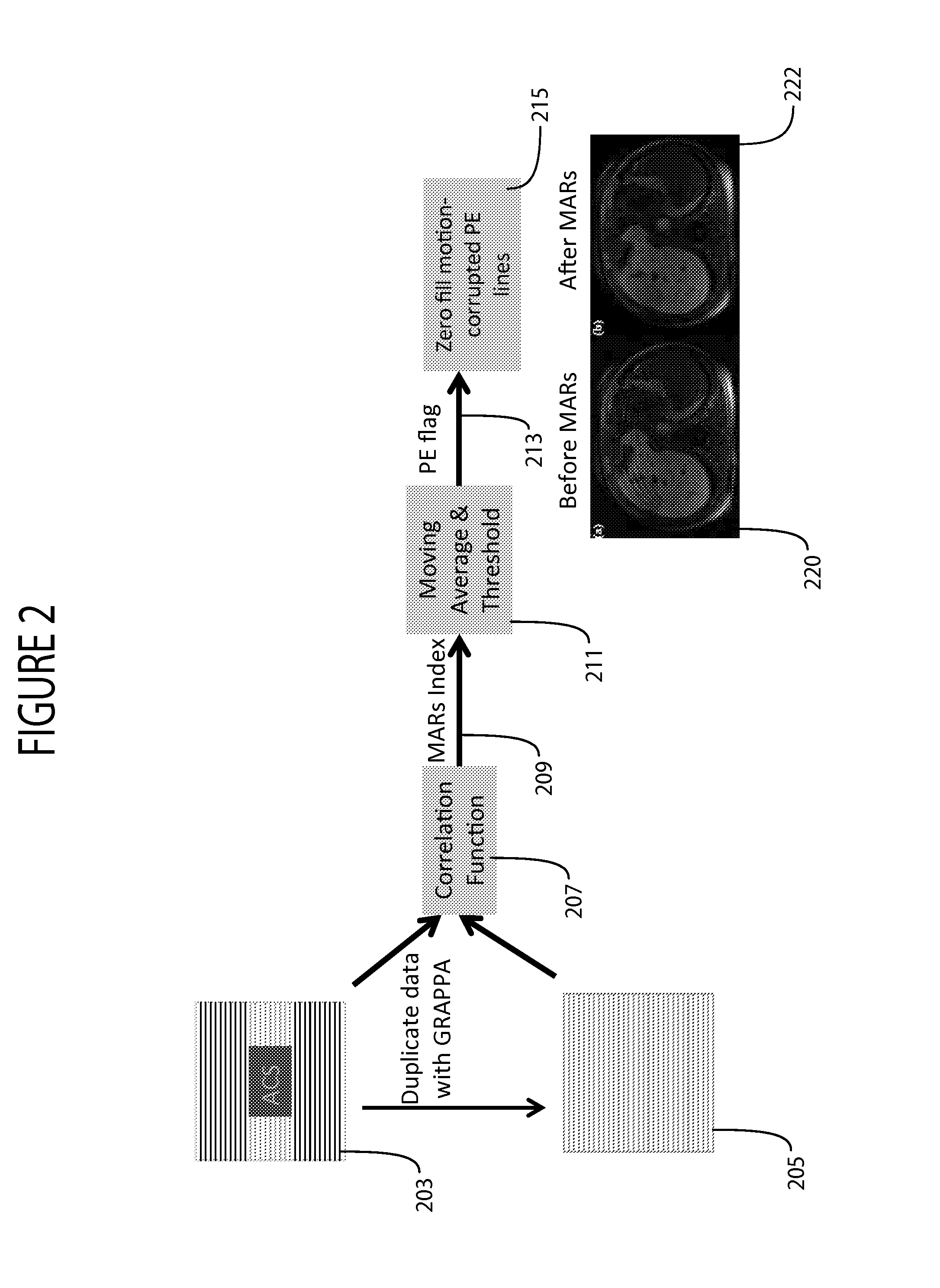
[0015]A system addresses respiratory related motion artifacts in medical imaging by exploiting measured information while minimizing need for patient breathhold cooperation and without substantially increasing image processing time, in clinical abdominal MR applications, for example. In discarding motion contaminated image data, it is desired to keep as much relevant information as possible. Known systems discard relevant image information and use just part of the acquired image data in employing sub-optimal methods to reduce respiratory motion artifacts. For instance, known methods typically use a gating method without employing fast acquisition methods or during patient breathhold use a fast acquisition without using gating information and discard useful image information.

[0016]A system according to invention principles uses breathhold, center-out k-space encoding and advantageously selects and discards motion contaminated data in a postprocessing stage comprehensively based on av...


PUM


Abstract

A method provides motion corrected MR image data in an MR imaging system. The method employs an imaging method for acquiring k-space data of a k-space data array during an imaging scan and the k-space data represents image data of a patient anatomical region. The method acquires k-space data associated with a central region of the k-space data array and subsequently acquires k-space data external to the central region of the k-space data array. The method acquires a motion signal indicating respiratory motion at least during the acquisition of the k-space data external to the central region and compares the motion signal with a predetermined threshold. The method identifies acquired k-space data corresponding to acquisition periods when the motion signal exceeds the threshold and excludes use of the identified acquired k-space data in image reconstruction using the remaining acquired k-space data.

Description

CROSS-REFERENCE TO RELATED APPLICATIONS[0001]This application is a non-provisional of, and claims priority to, U.S. provisional patent application Ser. No. 61 / 613,006 by K. Liu et al. filed on Mar. 20, 2012.FIELD OF THE INVENTION[0002]This invention concerns a system for providing motion corrected MR image data in an MR imaging system by excluding use of acquired k-space data in image reconstruction that is acquired in acquisition periods when a respiratory motion indicative signal exceeds a threshold.BACKGROUND OF THE INVENTION[0003]MRI body or body relevant region image quality is affected by patient respiration motion. Respiration related MR imaging artifacts occur if they are not suppressed or reduced, which leads to ghosting that may overlay a region of interest or blurring which reduces image quality for clinical diagnosis. Respiration related patient physical motion is unavoidable during MR imaging, especially for body region imaging, such as of the abdomen, spine, pelvis and...

Claims


Application Information

Patent Timeline
Patent Type & Authority Applications(United States)
IPC IPC(8): G06K9/36
CPCG01R33/5673
Inventor LIU, KECHENGZHAO, TIEJUN
Owner SIEMENS HEALTHCARE GMBH
Why Eureka
Try Eureka