Medical Instrument For Cutting Off Tissue And Cartilage From A Human Or Animal Body

a technology of medical instruments and cartilage, applied in the field of medical instruments, can solve the problems of difficult to avoid so-called debridement, large diameter, and inability to achieve the effect of easy sucking

Active Publication Date: 2013-10-17
KARL STORZ GMBH & CO KG
View PDF6 Cites 11 Cited by
  • Summary
  • Abstract
  • Description
  • Claims
  • Application Information

AI Technical Summary

Benefits of technology

[0034]This measure, likewise known per se, has the advantage that the separated pieces of connective tissue and also the pieces of cartilage separated by the curette can be fragmented into very small parts by the plurality of windows, which can optionally also have different sizes and geometries, and these very small parts can then be easily sucked out through the inner shaft. In this way, the risk of the inner shaft becoming blocked by pieces of tissue or cartilage is eliminated.
[0035]It has proven particularly advantageous if three such openings are present in the inner shaft.

Problems solved by technology

It is hard to avoid so-called debridement, i.e. where separated pieces of cartilage are released into the joint, which then necessitates irrigation of the joint in order to remove the small pieces, called chips.
However, the pieces of cartilage separated by the curette then always have to be smaller than the suction cross section of the instrument, since otherwise the suction cross section is blocked by the pieces of cartilage.
Accordingly, the suction channel would then need to have a correspondingly large diameter, which is not feasible in defect sites with diameters of a few centimetres, since such a large amount of space is unavailable in operations on joints.

Method used


Image

  • Medical Instrument For Cutting Off Tissue And Cartilage From A Human Or Animal Body
  • Medical Instrument For Cutting Off Tissue And Cartilage From A Human Or Animal Body
  • Medical Instrument For Cutting Off Tissue And Cartilage From A Human Or Animal Body

Examples


Embodiment Construction

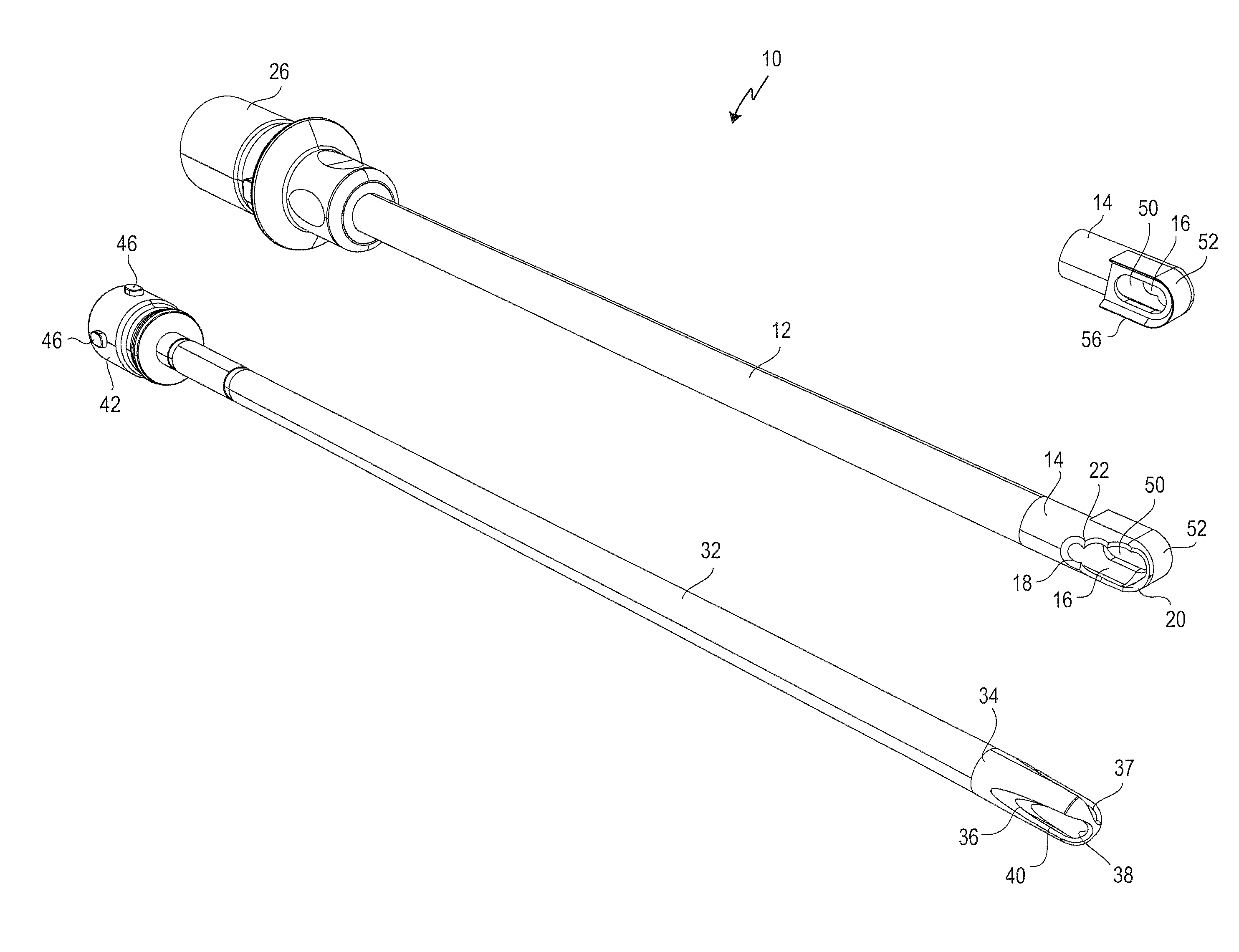
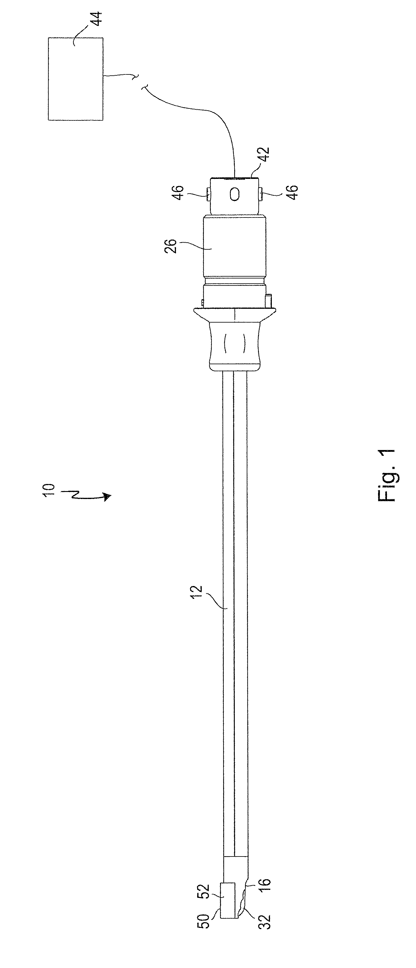
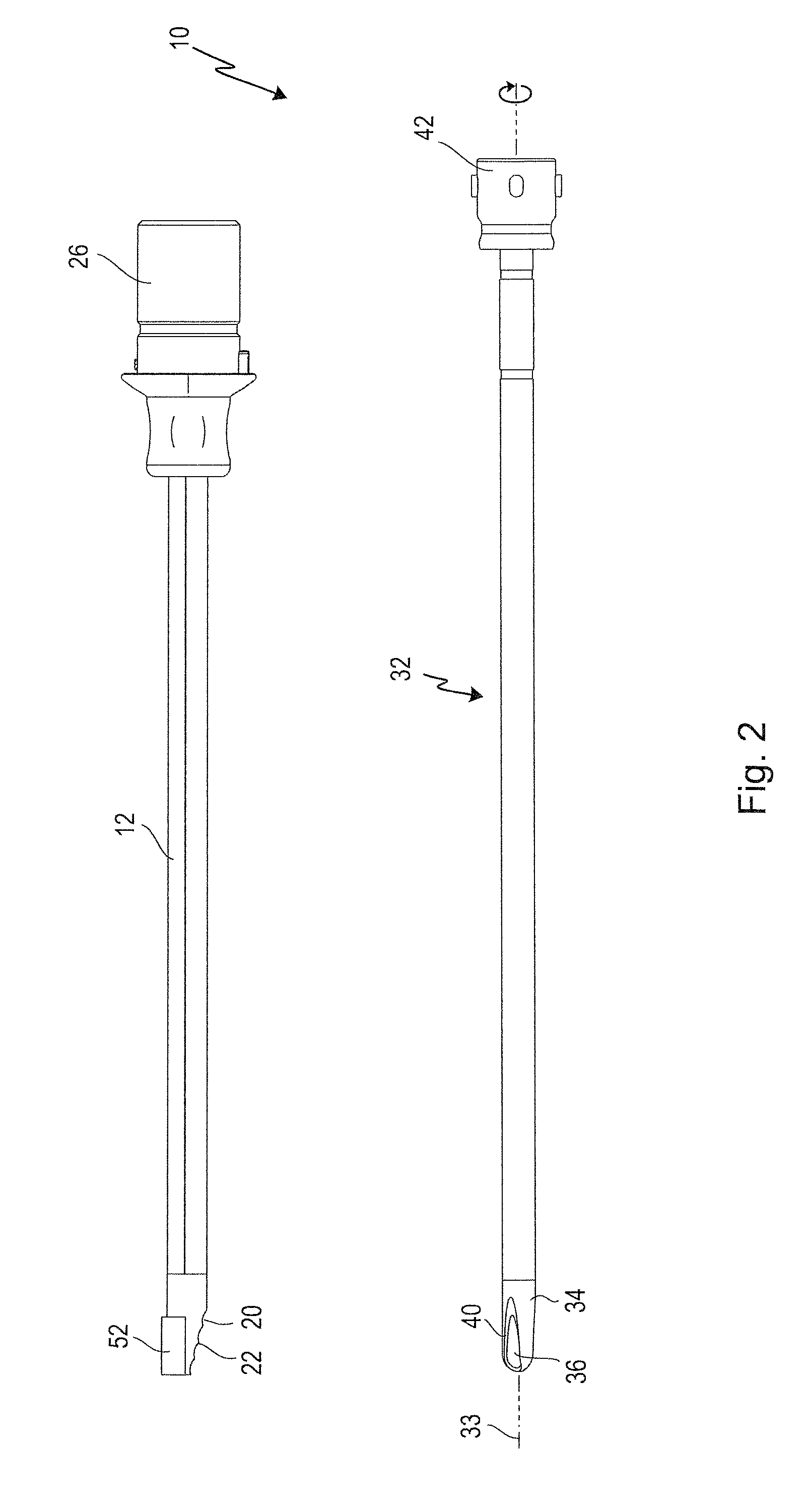
[0055]A medical instrument according to the invention shown in FIGS. 1 to 7, and used for separating tissue and cartilage, is designated in its entirety by the reference number 10.

[0056]The instrument 10 has an elongate, rectilinear hollow outer shaft 12.

[0057]The latter has, in the area of its distal end 14, a first window 16, as can be seen particularly from FIG. 3.

[0058]The single first window 16 has a circumferential border 18, which is designed as a cutting edge 20.

[0059]Teeth 22 stand vertically from the cutting edge 20, as can be seen particularly from FIGS. 3, 4 and 5.

[0060]The circumferential border 18 lies in the area of an oblique cut 24, as is indicated in FIG. 4. This oblique cut 24 is inclined radially inwards, as seen from proximal to distal, such that the first window 16 has an approximately oval cross section. As can be seen from FIG. 4, the inclination of the oblique cut 24 is such that less than half a diameter is cut away at the distal apex.

[0061]The teeth 22 can...


PUM


Abstract

A medical instrument serves for cutting off tissue and cartilage from a body. It comprises an outer shaft having at least a first window in a distal end area thereof. A hollow inner shaft is received within the outer shaft and can be rotated about a longitudinal axis of the outer shaft. The hollow inner shaft has at least one opening cooperating with the at least one window of the outer shaft in a cutting action when turning the inner hollow shaft. A further window is provided in the distal end area of the outer shaft which further window is surrounded by a curette having a separating edge. Tissue and cartilage separated by the curette can be cut into small pieces by the cutting edges of the rotatable inner shaft and then be sucked in through an opening of the inner hollow shaft.

Description

BACKGROUND OF THE INVENTION[0001]The invention relates to a medical instrument for separating tissue and cartilage from a body.[0002]One kind of such instruments, so-called shaver blades, is known from U.S. Pat. No. 4,811,734. For this purpose, the distal end of the outer shaft is guided to the operating site where the tissue that is to be separated is located. To separate the tissue, the inner shaft is moved in rotation by means of an external or internal drive. As they rotate, the cutting edges formed on the inner shaft cooperate in a cutting action with a border, designed as a cutting edge, of a window of the outer shaft. The cutting edge of an opening of the inner shaft runs past the cutting edge of the window in the outer shaft on each rotation. In order to ensure that the tissue to be separated is brought between the interacting cutting edges, the inner shaft can be connected to a vacuum source whose suction effect reaches through the inner shaft as far as the window on the ou...

Claims


Application Information

Patent Timeline
Patent Type & Authority Applications(United States)
IPC IPC(8): A61B17/16
CPCA61B17/16A61B17/1659A61B17/32002A61B17/320708A61B2017/00353A61B10/0266A61B17/320758A61B2017/320775A61B17/1604A61B2017/320024
Inventor STEINWACHS, MATTHIASBERBERICH, SASCHA
Owner KARL STORZ GMBH & CO KG
Why Eureka
Try Eureka