Eureka AIR delivers breakthrough ideas for toughest innovation challenges, trusted by R&D personnel around the world.

Tissue resection device and related methods of use

Active Publication Date: 2013-12-12
BOSTON SCI SCIMED INC
View PDF17 Cites 15 Cited by
  • Summary
  • Abstract
  • Description
  • Claims
  • Application Information

AI Technical Summary

Benefits of technology

The present disclosure provides a medical device for resecting undesired mass from a patient's body using a minimally invasive surgical method. The device includes an elongated member with a proximal end, a distal end, and at least one channel extending between the two ends. The distal portion of the channel is displaced relative to the longitudinal axis of the elongated member and is defined by a cap secured to the distal end of the elongated member. The cap may be removably coupled to the elongated member and may include a wall with an open section proximal to the distal end of the cap. The device may also include a diverter tube within the cap and a flared opening in communication with the lumen of the elongated member. The method involves inserting the resection device into a body cavity and advancing an instrument through the elongated member to excise tissue. The technical effects of the invention include minimizing damage to the patient's body and reducing the risk of complications.

Problems solved by technology

This procedure, however, may cause discomfort to patients, and pose health risks.
It is often difficult to grasp these polyps, without drawing in a part of the muscularis layer.
Because polyps or lesions are present on the organ walls, it is often difficult to grasp such objects readily with axially extending devices and obtain a better vantage point to apply overhead lifting force.

Method used


Image

  • Tissue resection device and related methods of use
  • Tissue resection device and related methods of use
  • Tissue resection device and related methods of use

Examples


Embodiment Construction

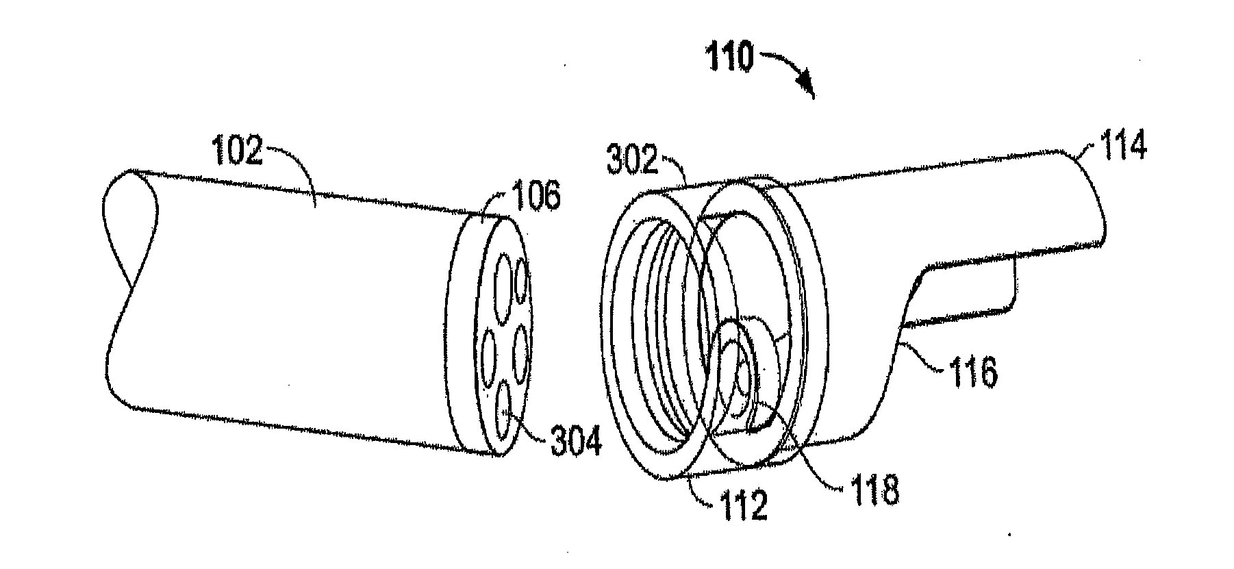
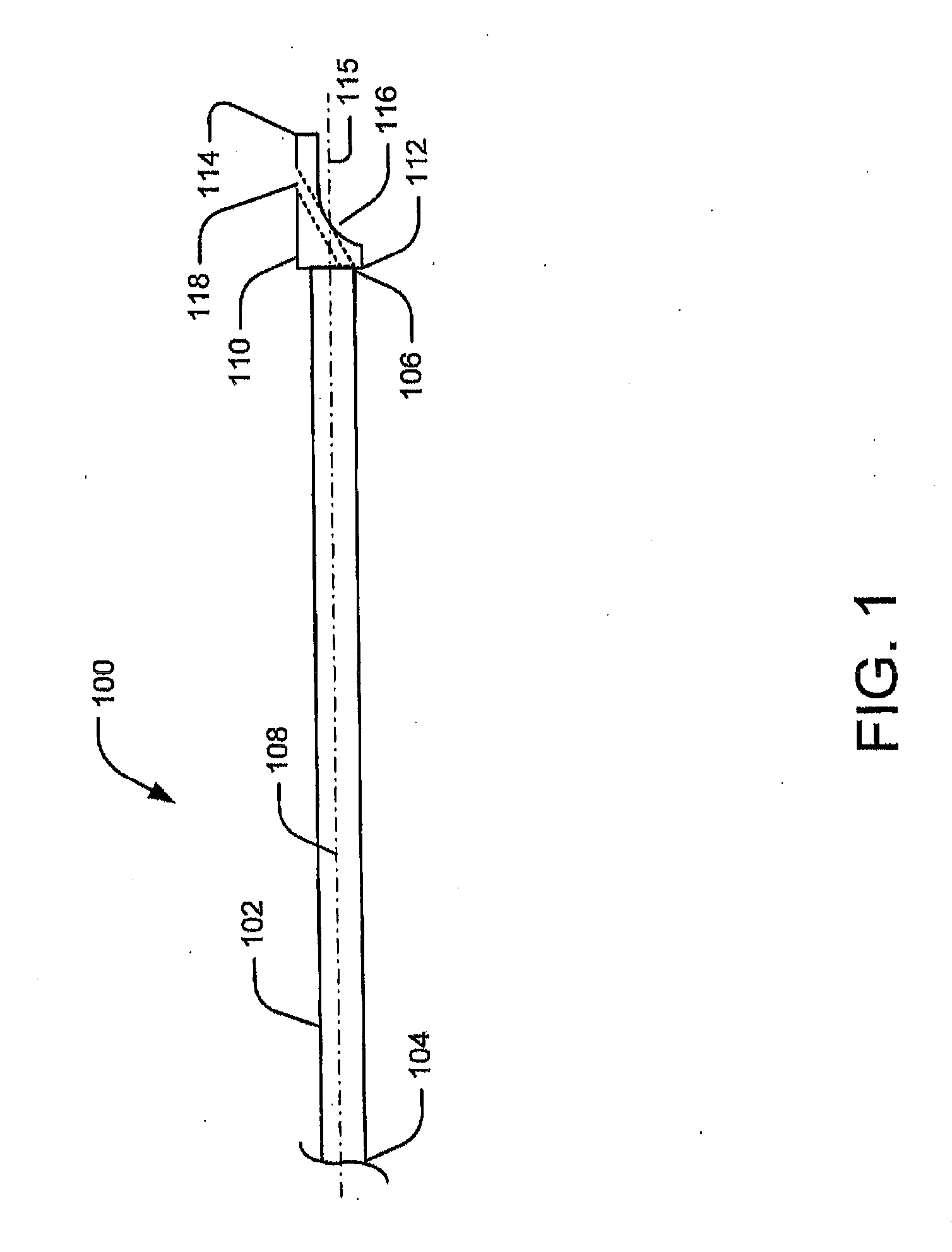
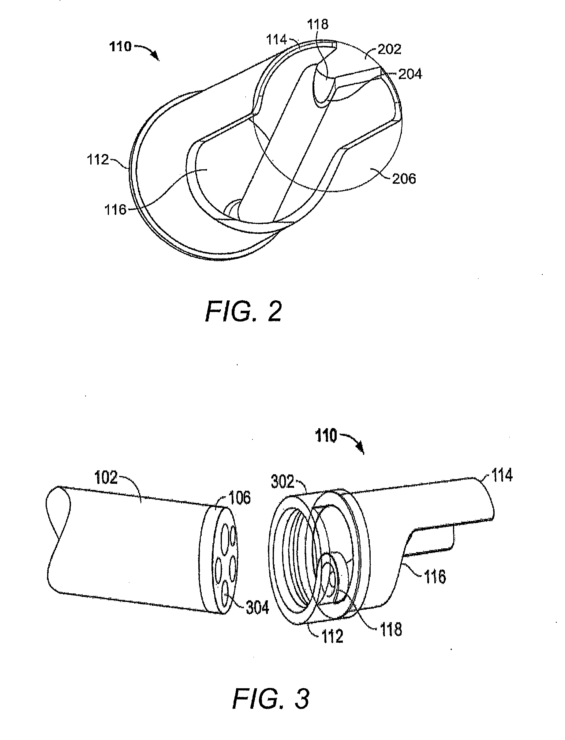
[0023]Reference will now be made in detail to embodiments of the present disclosure, an example of which is illustrated in the accompanying drawings. Wherever possible, the same reference numbers will be used throughout the drawings to refer to the same or like parts. The term “distal” refers to the end farthest away from a medical professional when introducing a device in a patient. By contrast, “proximal” refers to the end closest to the medical professional when placing a device in the patient.

Overview

[0024]Embodiments of the present disclosure relate to devices and methods for resecting or banding cancerous or otherwise undesirable tissue in a patient's body. For example, the device may be used in a minimally invasive procedure to remove cancerous polyps or lesions from the mucosal walls of the colon, esophagus, stomach, duodenum, or any other suitable location. A physician may also wish to resect tissue to conduct a biopsy or other examination. It should be understood that the ...


PUM


Abstract

Embodiments of the present disclosure include a medical device including, among other things, an elongated member having a proximal end, a distal end, and at least one channel. A distal portion of the at least one channel may be displaced relative to a longitudinal axis of the elongated.

Description

CROSS-REFERENCE TO RELATED APPLICATIONS[0001]This application claims the benefit of priority from U.S. Provisional Application No. 61 / 658,700 filed on Jun. 12, 2012, the entirety of which is incorporated by reference herein.FIELD OF THE INVENTION[0002]This disclosure relates generally to devices and methods for resecting tissue. More particularly, embodiments of the disclosure relate to minimally invasive devices and methods for endoscopic mucosal resection and endoscopic submucosal dissection.BACKGROUND OF THE INVENTION[0003]Organ walls are composed of several layers: the mucosa (the surface layer), the submucosa, the muscularis (muscle layer), and the serosa (connective tissue layer). In gastrointestinal, colonic, and esophageal cancer, for example, small lesions or cancerous masses may form along the mucosa and often extend into the lumens of the organs. Conventionally, that condition is treated by cutting out a portion of the affected organ wall. This procedure, however, may cau...

Claims


Application Information

Patent Timeline
IPC IPC(8): A61B17/221
CPCA61B17/221A61B2017/00269A61B2017/00296A61B17/00234A61B17/12013A61B2017/3445
Inventor SMITH, PAULSUON, NAROUNCARRILLO, JR., OSCAR R.RAYBIN, SAMUELWEITZNER, BARRY
Owner BOSTON SCI SCIMED INC
Why Eureka
Eureka Blog
Learn More