

Internal focus reference beads for imaging cytometry
- Summary
- Abstract
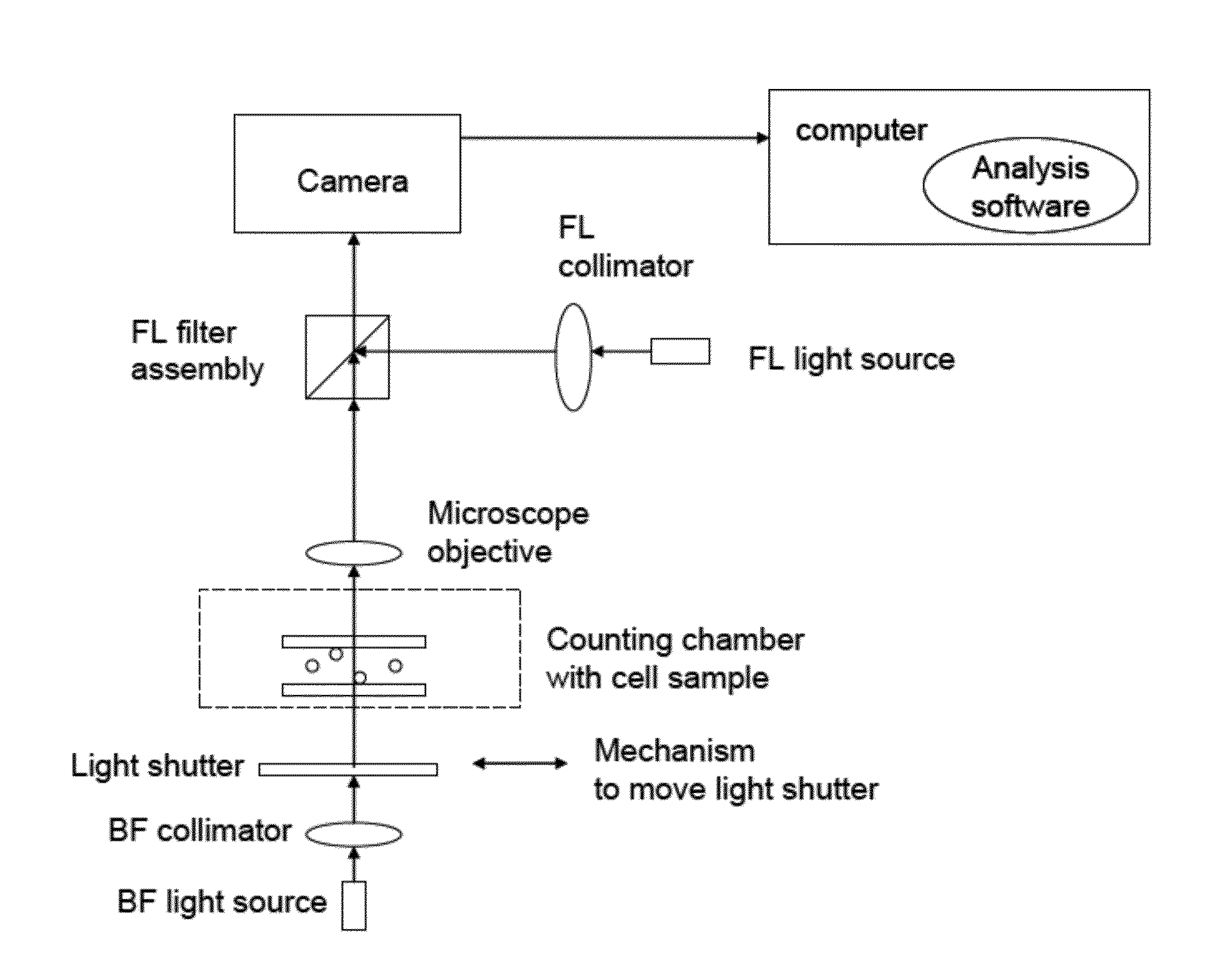
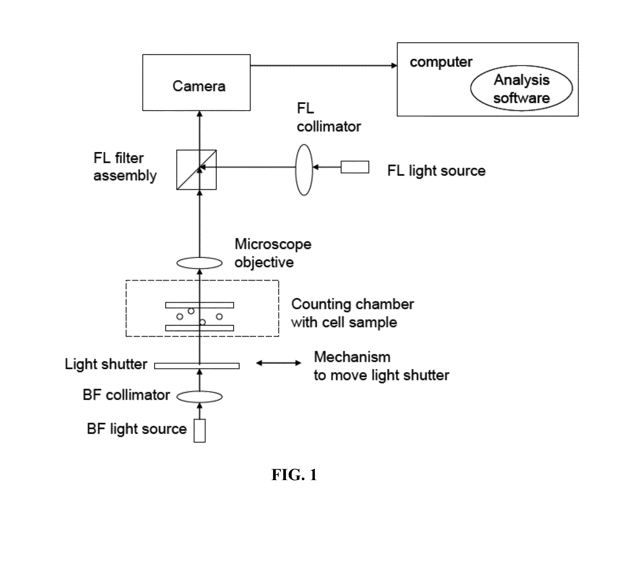
- Description
- Claims
- Application Information
AI Technical Summary
Benefits of technology
Problems solved by technology
Method used
Image


Examples
example 1
Imaging of Leukocytes
[0051]Using a large format camera, a large volume of blood was imaged, specifically for counting leukocytes for validating leukoreduced blood sample. Two images were captured (FIG. 6), one with excitation of UV and other with blue light. The blood sample was diluted 1 to 1 with Acridine Orange stain and fluorescent beads that fluoresces in both channels.
[0052]Counting only the beads in the UV excitation channel and counting all the particles in the blue excitation channel (beads+AO stained leukocytes), a subtraction result in a measurement of the leukocyte concentration in the blood sample.
example 2
Imaging of Leukocytes
[0053]Using PI stained jurkat, with optical modules QMAX blue with 510 nm long pass filter, and 535 nm-401 nm with 510 nm long pass filter, two images were taken with UV and blue excitation (FIG. 7 and FIG. 8).
[0054]No fluorescence of beads was observed in the channel that detects PI stained Jurkats. Method would involve focusing under UV excitation, with beads, then switching to blue excitation and counting all the Cells stained with PI. This is a much simpler method than the previous described method, where a subtraction is not needed. The method is using one channel with beads to focus, and then switch to the other channel to count all the cells.
PUM
 Abstract
Description
Claims
Application Information