Intra-oral x-ray imaging device equipped with camera

Active Publication Date: 2014-02-20
REMEDI CO LTD
View PDF10 Cites 8 Cited by
  • Summary
  • Abstract
  • Description
  • Claims
  • Application Information

AI Technical Summary

Benefits of technology

[0035]As described above, in an intraoral X-ray imaging apparatus having a camera according to the present invention, since an X-ray irradiator is located inside the oral cavity, it is possible to obtain an image having a high quality with a low X-ray dose and to avoid confusion in image diagnosis by removing a ghost image.
[0036]In addition, since the imaging object tooth can be accurately identified inside the oral cavity, the X-ray can be irradiated on only the imaging object tooth, so that it is possible to minimize a radiation exposure dose applied to the head of a patient.
[0037

Problems solved by technology

Accordingly, the X-ray imaging apparatus in the related art has problems as follows.
The first problem is that unnecessary radiation exposure dose may be applied to the head.
The second problem is that a ghost image, that is, an image of a tooth other than the tooth of interest may exist.
The third problem is that images of a tooth captured at various angles cannot be obtained.
The fourth problem is that the X-ray imaging apparatus occupies a too large space.
However, in

Method used


Image

  • Intra-oral x-ray imaging device equipped with camera
  • Intra-oral x-ray imaging device equipped with camera
  • Intra-oral x-ray imaging device equipped with camera

Examples


Example

BEST MODE

[0050]Hereinafter, an intraoral X-ray imaging apparatus having a camera according to a preferred embodiment of the present invention will be described in detail with reference to the attached drawings.

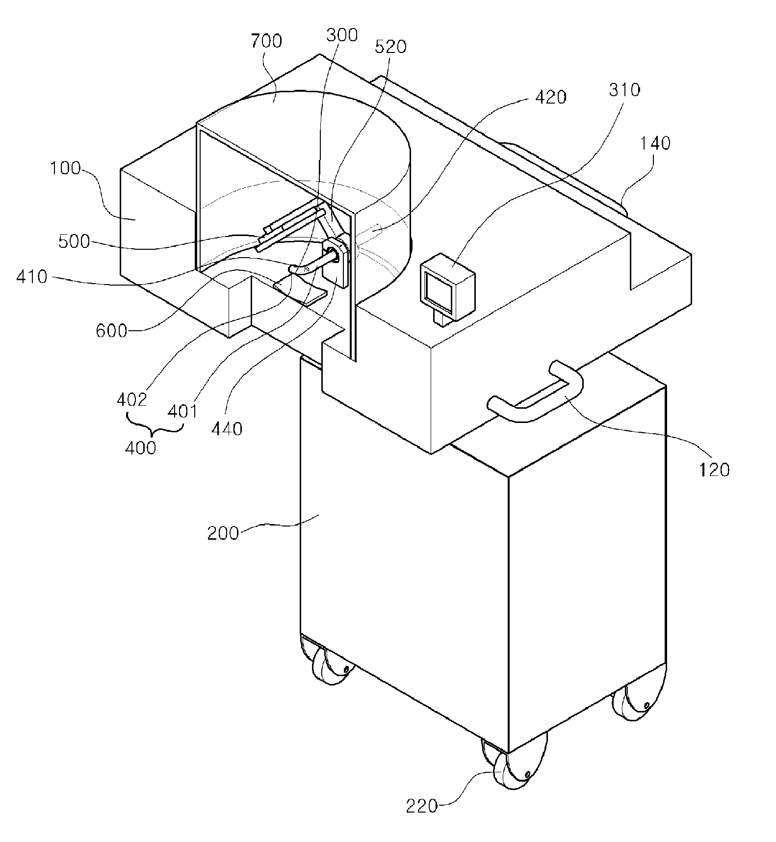
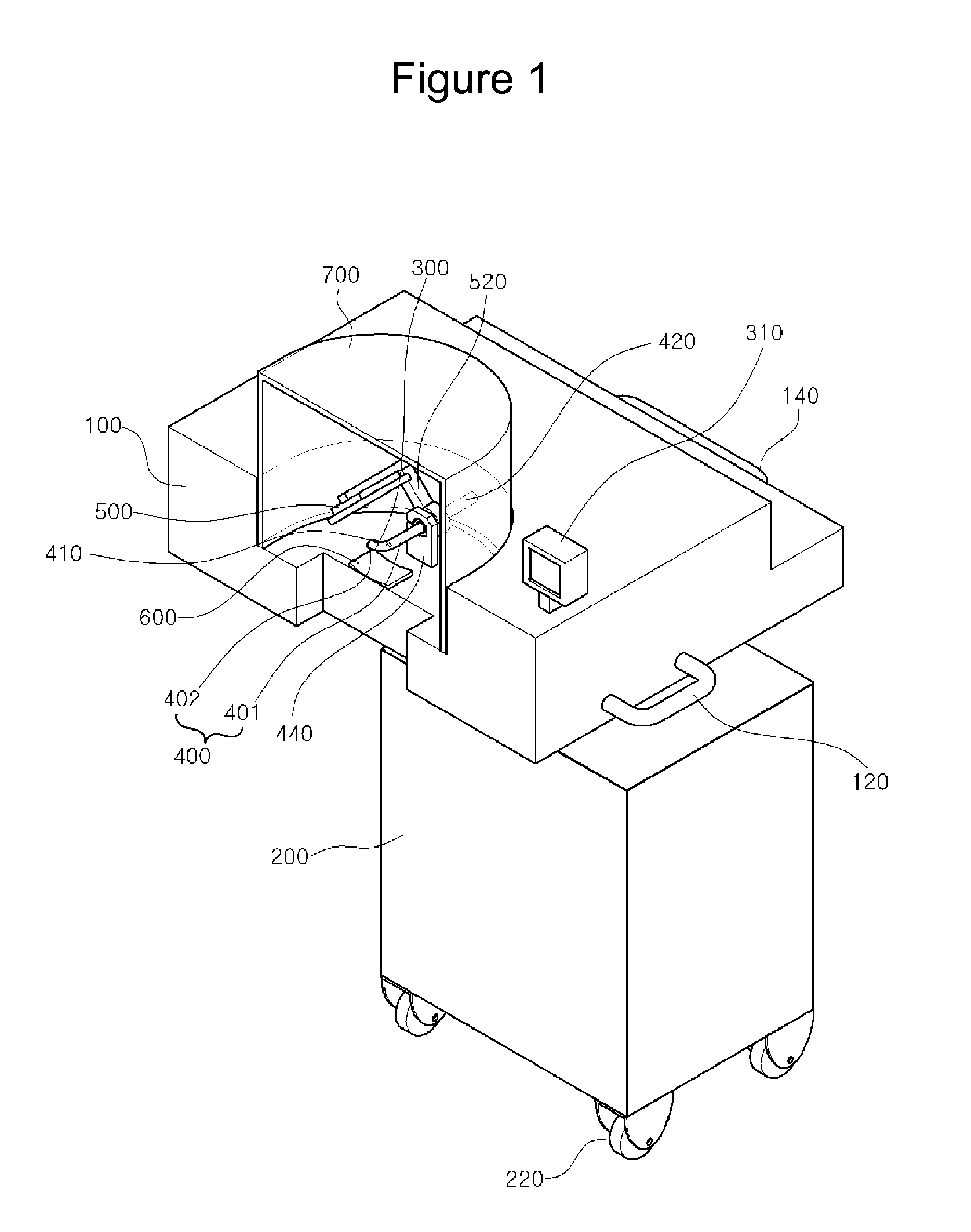
[0051]FIG. 1 illustrates an intraoral X-ray imaging apparatus having a camera according to an embodiment of the present invention.

[0052]The intraoral X-ray imaging apparatus having a camera according to the present invention is configured to include a frame 100.

[0053]The frame 100 is provided with a grip 120, so that a patent can fix the pose by holding the grip 120.

[0054]The frame 100 is provided with a shield glass 700, so that leakage of X-ray irradiated from the X-ray irradiator 400 can be prevented. Therefore, it is possible to prevent persons other than the patient from being exposed to radiation.

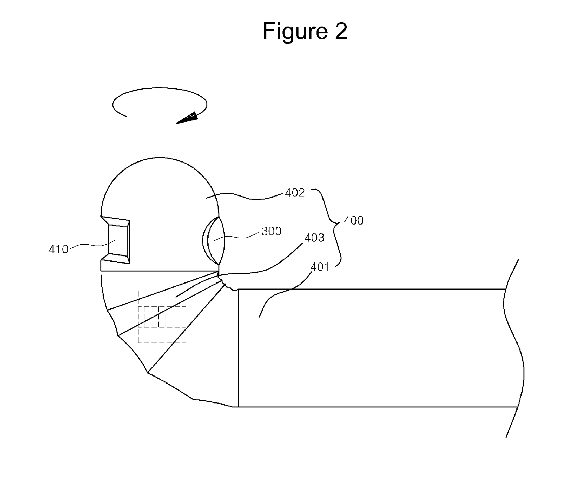
[0055]The intraoral X-ray imaging apparatus having a camera according to the present invention is configured to an X-ray irradiator 400 which is installed so as to be supported ...


PUM


Abstract

Provided is an intraoral X-ray imaging apparatus having a camera, including: a frame; an X-ray irradiator which is installed to be supported by the frame and of which the one end portion is inserted into the oral cavity and which irradiates X-ray through an X-ray irradiation hole formed on the one end portion inserted into the oral cavity; a camera unit which is installed at the one end portion of the X-ray irradiator to image an interior of the oral cavity; a monitor unit which is connected to a camera unit to output the image captured by the camera unit; and an X-ray detector which is arranged to be separated from the X-ray irradiator in the frame so as to detect the X-ray irradiated from the X-ray irradiator outside the oral cavity. In the intraoral X-ray imaging apparatus having a camera, since the X-ray irradiator is located inside the oral cavity, it is possible to obtain an image having a high quality with a low X-ray dose and to avoid confusion in image diagnosis by removing a ghost image. In addition, since the imaging object tooth can be accurately identified inside the oral cavity, the X-ray can be irradiated on only the imaging object tooth, so that it is possible to minimize a radiation exposure dose applied to the head of a patient.

Description

TECHNICAL FIELD[0001]The present invention relates to an intraoral X-ray imaging apparatus having a camera, and more particularly, to an intraoral X-ray imaging apparatus having a camera capable of obtaining an accurate image having constant magnification in the case of obtaining a panoramic image for dental treatment by using an X-ray irradiator with a low X-ray dose located inside an oral cavity.BACKGROUND ART[0002]In general, an X-ray imaging apparatus for identifying a state of an alveolar bone (teeth-associated structure) including teeth for teeth / periodontal disease treatment and orthodontic treatment is installed in a dental clinic.[0003]An operator (or a dentist) uses the X-ray imaging apparatus to identify the size and shape of the jawbone and an anatomical change of the jawbone in the horizontal and vertical axes through radiological tests and to consider disease factors existing in the jawbone and information influencing medical treatment. The image information is importa...

Claims


Application Information

Patent Timeline
IPC IPC(8): A61B6/14
CPCA61B6/145A61B6/4405A61B6/4429A61B6/14
Inventor LEE, RENA
Owner REMEDI CO LTD
Why Eureka
Try Eureka