Open surgery anastomosis device, system, and method

a technology of anastomosis device and open surgery, applied in the field of open surgery anastomosis device, system and method, can solve the problems of complex and delicate suturing techniques, unsatisfactory and unsuccessful suturing, and suturing techniques that require a very high level of skill

Inactive Publication Date: 2014-02-20
SINGH DEVINDER +1
View PDF9 Cites 31 Cited by
  • Summary
  • Abstract
  • Description
  • Claims
  • Application Information

AI Technical Summary

Benefits of technology

[0014]In another embodiment, the medical device system includes a first external structure and a second external structure each sized and configured to be positioned over separate portions of the tubular structure with me anatomical structure therebetween. Such first and second external structures may be employed to further prevent potential leaks and migration of the anatomical structure from the tubular structure.
[0017]In another embodiment, the method step of inflating the balloon includes forcing fluid into the balloon through a balloon port oriented substantially transverse relative to the longitudinal length of the tabular structure and positioned within the port extension. In yet another embodiment, the method step of inflating the balloon includes radially expanding the tubular structure having multiple tines extending therefrom so that the tubular structure enlarges to a size that substantially prevents migration of the first and second ends of the hollow anatomical structure from the tubular structure to, thereby, provide a fluid flow path through the tubular structure.

Problems solved by technology

However, such process is known to be time consuming and, due to the size of some vessels, such suturing often is unsatisfactory and unsuccessful.
This is especially true for the anastomosis of vessels in micro-surgical procedures that require more complex and delicate suturing techniques.
When dealing with small vessels, the challenge is to establish reliable fluid flow using microvascular suture techniques that provide for the anastomosis without constricting the vessels and also preventing leaks from the anastomosis or repair site.
Such technically complex micro-vascular suturing techniques require a very high level of skill, and nevertheless a common occurrence is accidental capture of the vessel backwall in the suture, which then provides art unsatisfactory result in achieving a proper fluid flow path.
In sum, the complex procedures and suturing techniques necessary to employ proper anastomosis between various vessels is a time consuming process and often yields unsatisfactory results.
Further, the longer open surgery lasts for a patient, the higher the risk for complications to the patient.

Method used


Image

  • Open surgery anastomosis device, system, and method
  • Open surgery anastomosis device, system, and method
  • Open surgery anastomosis device, system, and method

Examples


Embodiment Construction

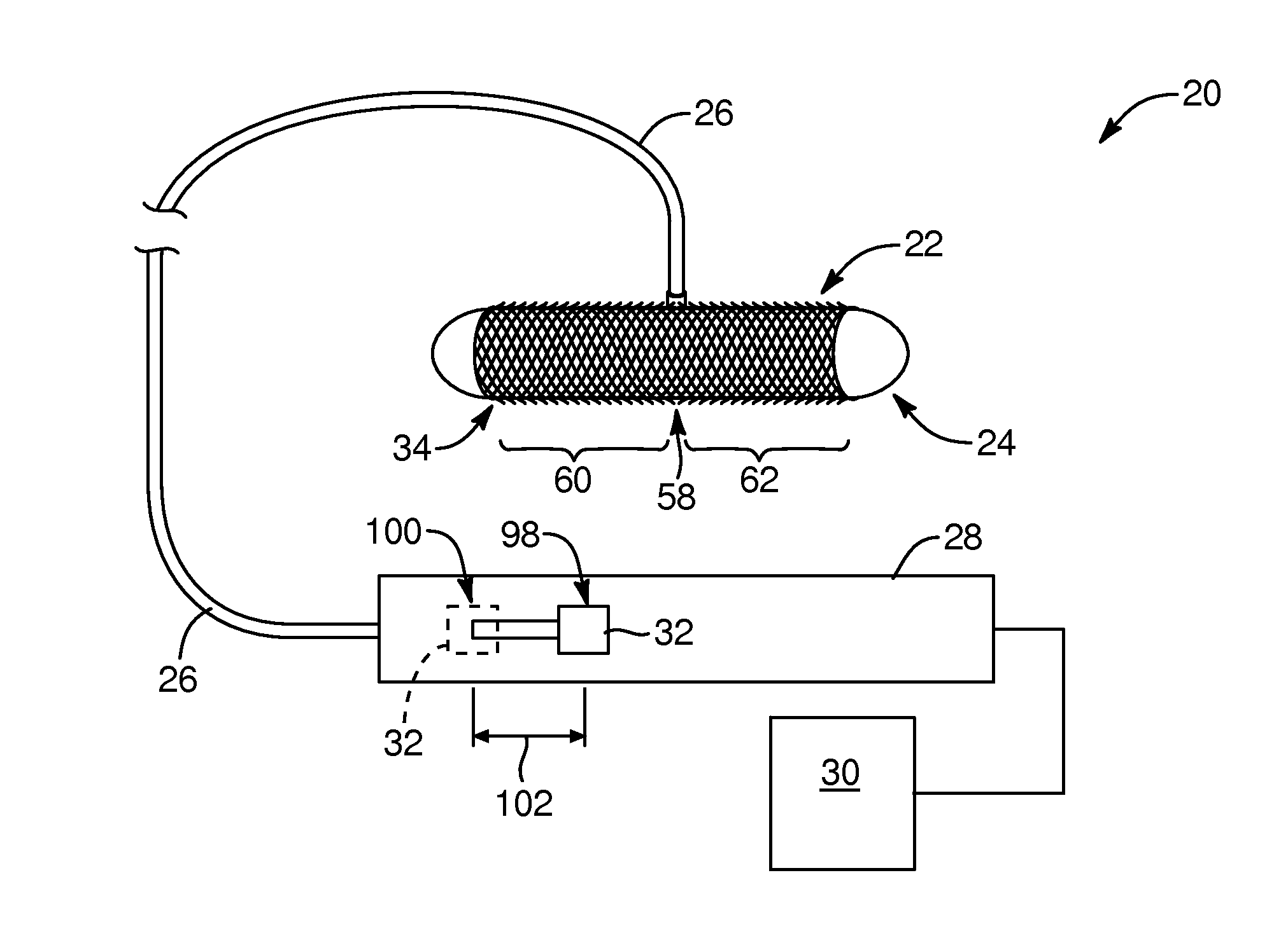
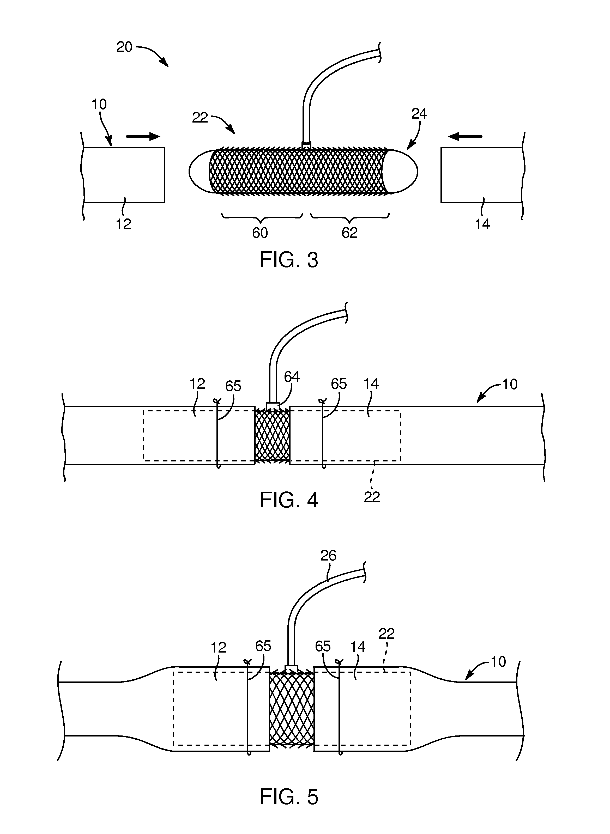
[0028]Referring to FIG. 1, a medical device system 20 configured to couple a tubular anatomical structure 10 (FIG. 3) is depicted. The medical device system 20 may include an anastomosis device 22, a balloon 24, and a fluid flow tube 26 extending between the balloon 24 and a handle 28. In addition, the handle 28 may be coupled to a fluid flow source 30. The handle 28 may include an actuator 32 configured to facilitate manual control of fluid flow between the fluid flow source and the balloon 24. The medical device system 20 of the present invention may be employed in open surgical, procedures to assist a physician in re-coupling, for example, severed (surgically divided or traumatically divided) hollow or tubular structures in the human anatomy, such as, arterial and venous vessels and / or lymphatic vessels. For example, the medical device system 20 may be employed for artery to artery coupling, vein to vein coupling, or artery to vein coupling (as well as vein to artery), or lymphat...


PUM


Abstract

A medical device, system and method tor an open surgical procedure configured to couple, for example, open ends of anatomical structures. The system includes an expandable tubular structure and inflatable balloon positioned within the tubular structure. The tubular structure includes a lateral opening defined in a wall of the tubular structure through which the balloon is inflated to radially expand the tubular structure, after which the balloon is deflated, and then removed from the tubular structure to, thereby, provide a fluid flow path through the tubular structure.

Description

CROSS-REFERENCE TO RELATED APPLICATIONS[0001]The present application claims the benefit of U.S. Provisional Application No. 61 / 684,111, filed on Aug. 16,2012, the disclosure of which is hereby incorporated by reference in its entirety.TECHNICAL FIELD[0002]The present invention relates generally to interconnecting tubular structures in the human anatomy in open surgical procedures and, more specifically, to anastomosis devices, systems, and methods for connecting blood vessels or other tubular structures in open surgical procedures.BACKGROUND[0003]Fluid-carrying tubular structures or vessels exist in a wide number of systems, including those physiological systems found in, for example, the human body. During an open surgical procedure it is frequently necessary to modify or repair various anatomical vessels that involve the connection or anastomosis of, for example, two ends to define a fluid path. Physicians typically have employed suturing techniques to connect the ends to establis...

Claims


Application Information

Patent Timeline
Patent Type & Authority Applications(United States)
IPC IPC(8): A61B17/11
CPCA61B17/11A61B2017/00022A61B2017/00557A61B2017/00893A61B2017/00995A61B2017/1107A61B2017/1132A61F2/064A61F2/958
Inventor SINGH, DEVINDERWHITE, JOSHUA J.
Owner SINGH DEVINDER
Why Eureka
Try Eureka