Three dimensional imaging data viewer and/or viewing

a three-dimensional imaging and data viewer technology, applied in the field of three-dimensional viewer for and/or three-dimensional viewing, can solve the problems of difficult viewing for viewers, difficult identification of depth locations, and much more difficult semi-transparent volume-rendered (vr) images

Inactive Publication Date: 2014-03-13
KONINKLIJKE PHILIPS ELECTRONICS NV
View PDF7 Cites 7 Cited by
  • Summary
  • Abstract
  • Description
  • Claims
  • Application Information

AI Technical Summary

Benefits of technology

[0009]In another aspect, a computer readable storage medium is encoded with one or more computer executable instructions, which, when executed by a proc...

Problems solved by technology

Unfortunately, when the 3D imaging data is visually presented on a 2D display monitor, it is often difficult for the viewer (i.e., the person visually observing the displayed data) to identify depth location of anatomical structure of interest.
While it may be relatively simple to “feel” the depth of a surface-shaded rendering (SSD), and even to make measurements on it, this becomes much more difficult on semi-transparent Volume-Rendered (VR) images.
Similarly, Maximum or Minimum Intensity Projection (MIP/MinIP) images, ...

Method used


Image

  • Three dimensional imaging data viewer and/or viewing
  • Three dimensional imaging data viewer and/or viewing
  • Three dimensional imaging data viewer and/or viewing

Examples


Embodiment Construction

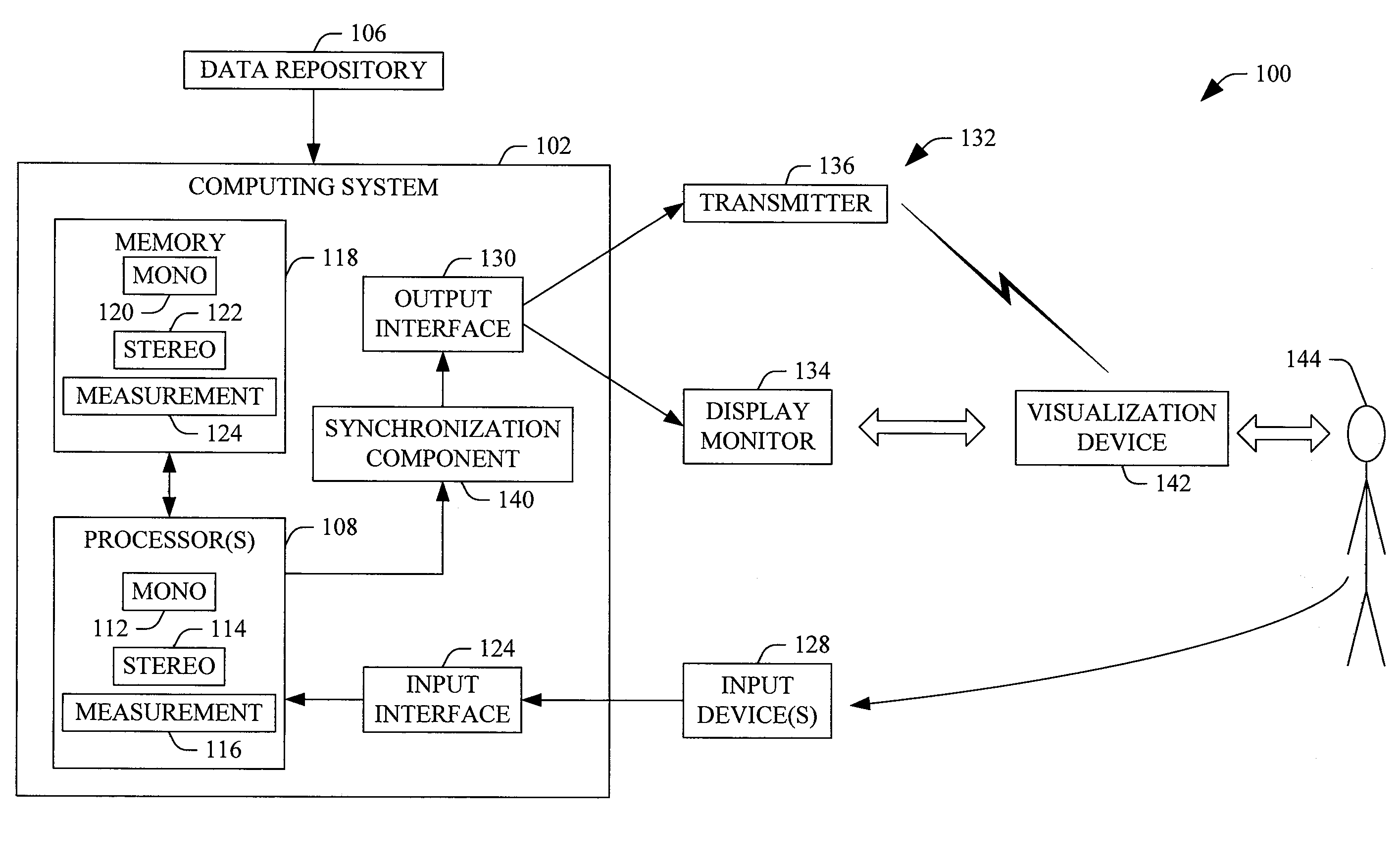
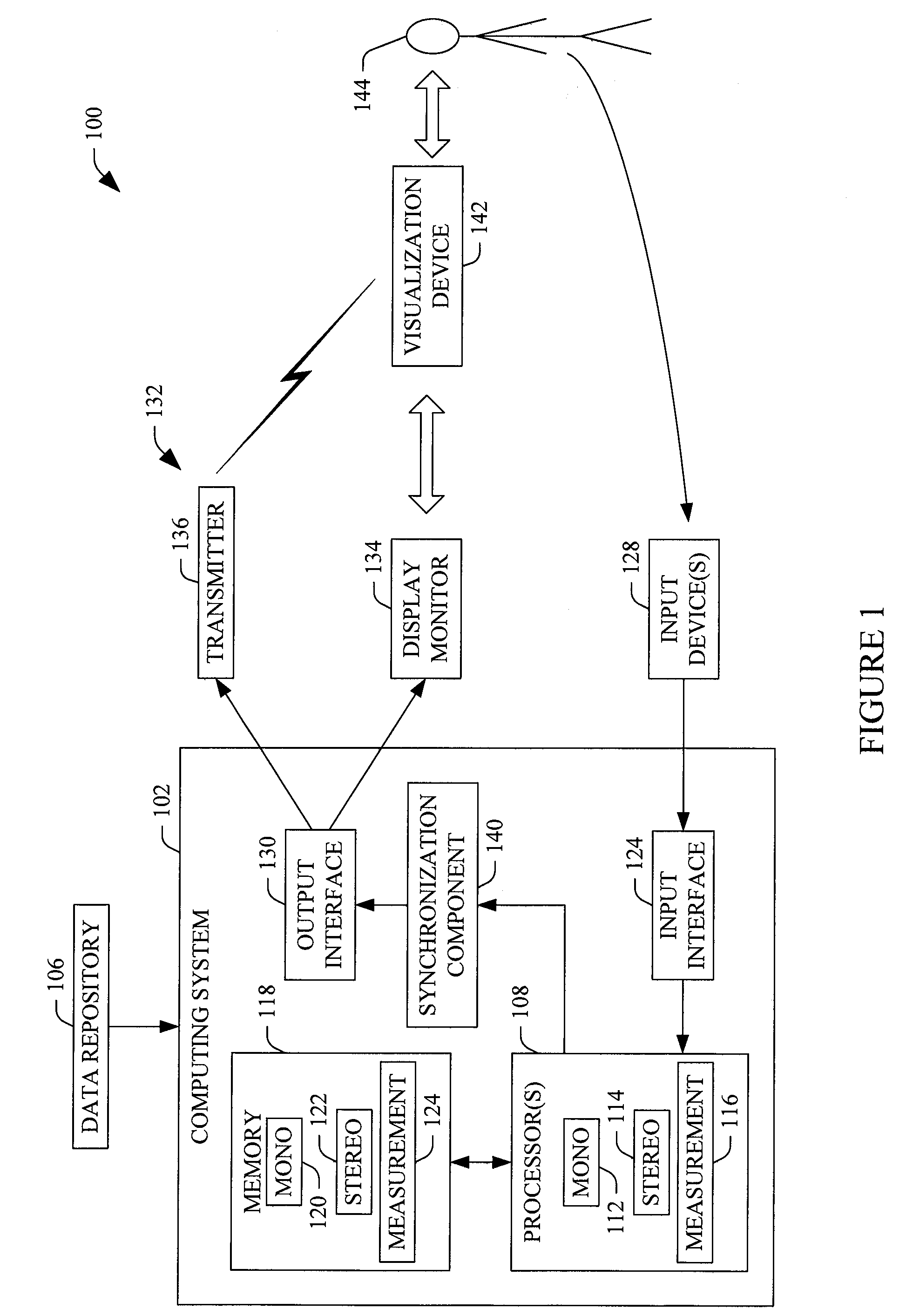
[0028]The following generally describes an approach in which imaging data (CT, MR, PET, etc.) is presented in 3D via a 2D display monitor by generating two images from two different viewpoints (e.g., left and right) that are shifted from each other by a predetermined distance (e.g., 10 mm) and / or angled by a predetermined angle (e.g., ±10 degrees), and visually presenting the two images stereoscopically. The values for the shift and angle can be default and / or user specified, and based on desired visible characteristics.

[0029]FIG. 1 illustrates a three dimensional viewing system 100. The system 100 includes a computing apparatus 102 that processes and visually presents three dimensional (3D) imaging data in three dimensions. Such data includes, but is not limited to, CT, MR, PET, etc. imaging data, which can be obtained from a data repository 106 such as a picture archiving and communication system (PACS), a radiology information system (RIS), a hospital information system (HIS), an...


PUM


Abstract

A method includes displaying three dimensional medical imaging data in three dimensions via a display monitor (134) by generating and visually presenting a stereoscopic view of the three dimensional medical imaging data in the display monitor. A system includes a stereo processor (114) that processes three dimensional medical imaging data and generates two images from two different viewpoints, which are shifted from each other by a predetermined distance and are angled by a predetermined angle, and a display monitor (134) used to alternately display the two images, thereby creating a stereoscopic view.

Description

FIELD OF THE INVENTION[0001]The following generally relates to a three dimensional viewer for and / or three dimensional viewing of imaging data generated by one or more imaging modalities such as computed tomography (CT), magnetic resonance (MR), positron emission tomography (PET), and / or other imaging modalities.BACKGROUND OF THE INVENTION[0002]Imaging modalities such as CT, MR, PET, etc. generate three dimensional (3D) imaging data indicative of a scanned object or subject. Unfortunately, when the 3D imaging data is visually presented on a 2D display monitor, it is often difficult for the viewer (i.e., the person visually observing the displayed data) to identify depth location of anatomical structure of interest.[0003]While it may be relatively simple to “feel” the depth of a surface-shaded rendering (SSD), and even to make measurements on it, this becomes much more difficult on semi-transparent Volume-Rendered (VR) images. Similarly, Maximum or Minimum Intensity Projection (MIP / M...

Claims


Application Information

Patent Timeline
IPC IPC(8): H04N13/04
CPCH04N13/0497H04N13/0486A61B6/02A61B6/466H04N13/279H04N13/117H04N13/128H04N13/315H04N13/398A61B6/022
Inventor GOTMAN, SHLOMO
Owner KONINKLIJKE PHILIPS ELECTRONICS NV
Why Eureka
Try Eureka