Methods and devices for endovascular therapy

a technology of endovascular therapy and endovascular valve, which is applied in the field of medical devices and methods, can solve the problems of limited long-term effectiveness of angioplasty and stent implantation, unsatisfactory methods and devices for delivering anti-stenotic therapy to blood vessel wall tissue, and far from solving restnosis problems, so as to reduce stenosis, inhibit restnosis, and increase vessel permeability.

Active Publication Date: 2014-05-22
CARDIOPROLIFIC INC
View PDF27 Cites 5 Cited by
  • Summary
  • Abstract
  • Description
  • Claims
  • Application Information

AI Technical Summary

Benefits of technology

[0050]Other embodiments of the present invention include devices and methods utilizing ultrasound energy propagated in the form of longitudinal waves, surface (radial or elliptic) waves and shear (transverse) waves (among other waves) to change compliance of plaque or vessel obstruction, and to increase vessel permeability.
[0051]Resistant fibrotic plaque or calcified plaque is a major limitation in treating arterial disease. Calcium deposits are frequent in patients with advanced age, diabetes, renal insufficiency, hypertension, and a history of smoking. Calcified lesions are challenging to treat with traditional endovascular therapy such as conventional angioplasty. Frequently, to displace the calcified plaque, balloon inflation pressures of more than 20 atm is required. Even after calcific plaque is disposed, and the vessel is open, it still continues to be a significant barrier for the delivery of therapeutic drug to the vessel wall to further reduce stenosis and inhibit restenosis. Ultrasound energy can erode and fracture calcified plaque while causing minimal injury to a healthy tissue. Application of ultrasound longitudinal waves, surface (radial or elliptic) waves and shear (transverse) waves may induce micro-cracks in the plaque and create micro-channels to further facilitate therapeutic drug penetration and permeability of the vessel.
[0052]In another embodiment, the invention provides devices and methods for ablating plaque, crossing Chronic Total Occlusions (CTO), as well as dissolving and removing blood clots and thrombus. This embodiment may include advancing a distal end of an ultrasound delivery device into the vessel, vein or other locations where plaque or blood clots are located, activating the ultrasound catheter to emit radial ultrasound energy, and applying aspiration or gravitational approach to further dissolve and remove blood clots outside the patient body. Use of therapeutic drug and / or microbubbles may enhance plaque and blood clots dissolving and removal process.
[0053]In patients undergoing endovascular procedures involving a vascular obstruction removal or administration of therapeutic drugs, use of ultrasound energy may be beneficial in the delivery of liquid medicament to further facilitate the distribution, delivery, absorption and / or efficacy of the medicament to improve clinical outcomes. Various ultrasonic catheter devices have been developed for use in ablating or otherwise removing obstructive material from blood vessels. For example, ultrasound devices with flexible ultrasound members for tissue ablation (either with or without therapeutic drugs) have been discussed in the prior art. Known devices enable translation of vibrations from the transducer to the flexible probe causing the probe to oscillate longitudinally or transversely. Longitudinal vibratory movement of the distal head or probe causes disintegration and ablation of the adjacent lesion while simultaneously delivering therapeutic drugs. Examples of such devices include U.S. Pat. Nos. 6,689,086 and 6,929,632 (both by Nita, et al). Other examples of devices utilizing longitudinal vibrations are described in U.S. Pat. Nos. 6,855,123; 6,942,677; 7,137,963; 7,297,131; 7,393,338; 7,621,929; 7,955,293; 8,133,236 and US Publication No. 2008 / 0108937 (all by Nita, et al.). Unlike longitudinal vibrations, transverse vibration emits a transverse ultrasonic energy along the length of the probe body so that a plurality of transverse nodes and anti-nodes are formed along the length of the probe. Examples of such devices include U.S. Pat. Nos. 6,524,251; 6,551,337; 6,652,547; 6,660,013; 6,695.781; 6,733,451; 6,866,670; 7,494,468; and 7,503,896 (all by Rabiner, et al.). While the longitudinal waves oscillate in the longitudinal direction or the direction of wave propagation, transverse waves oscillate perpendicular to the direction of propagation. Transverse waves are relatively weak compared to longitudinal waves and are known to not effectively propagate through liquids. Also, transverse vibrations are known to produce unwanted stress along the ultrasound transmission member, often causing breakage of the ultrasound transmission member. While longitudinal waves (as described in the prior art) are powerful, their ability to deliver ultrasound energy to the surrounding vessel wall is limited to the vibrating tip area. Therefore, longitudinal and transverse vibrations are limited in inducing uniform cellular changes along the treated vessel wall to further facilitate drug therapies.
[0054]The present invention provides methods and devices configured to deliver ultrasound surface waves or radial vibrational energy along an ultrasound transmission member to the surrounding tissue, to either small or large vessels or other cavities, and consequently increasing vessel drug uptake. Such devices provide a desired level of ultrasound energy to induce cellular changes while preventing vascular damage and reducing the potential of breakage for the ultrasound transmission member.

Problems solved by technology

Atherosclerosis and its consequences, including arterial stenosis, venous stenosis and hypertension, represent a major health problem both in the U.S. and throughout the world.
Angioplasty and stent implantation, however, often are of limited long term effectiveness due to restenosis and reocclusion.
While these approaches have met with some success, the restenosis problem is far from solved, as drug eluting stents and balloons have had mixed results in clinical studies.
While holding considerable promise, the methods and devices for delivering antistenotic therapeutic agents to blood vessel wall tissue are as yet not fully satisfactory.
Absorption of the therapeutic agent into the blood vessel wall, for example, represents a significant challenge.

Method used


Image

  • Methods and devices for endovascular therapy
  • Methods and devices for endovascular therapy
  • Methods and devices for endovascular therapy

Examples


Embodiment Construction

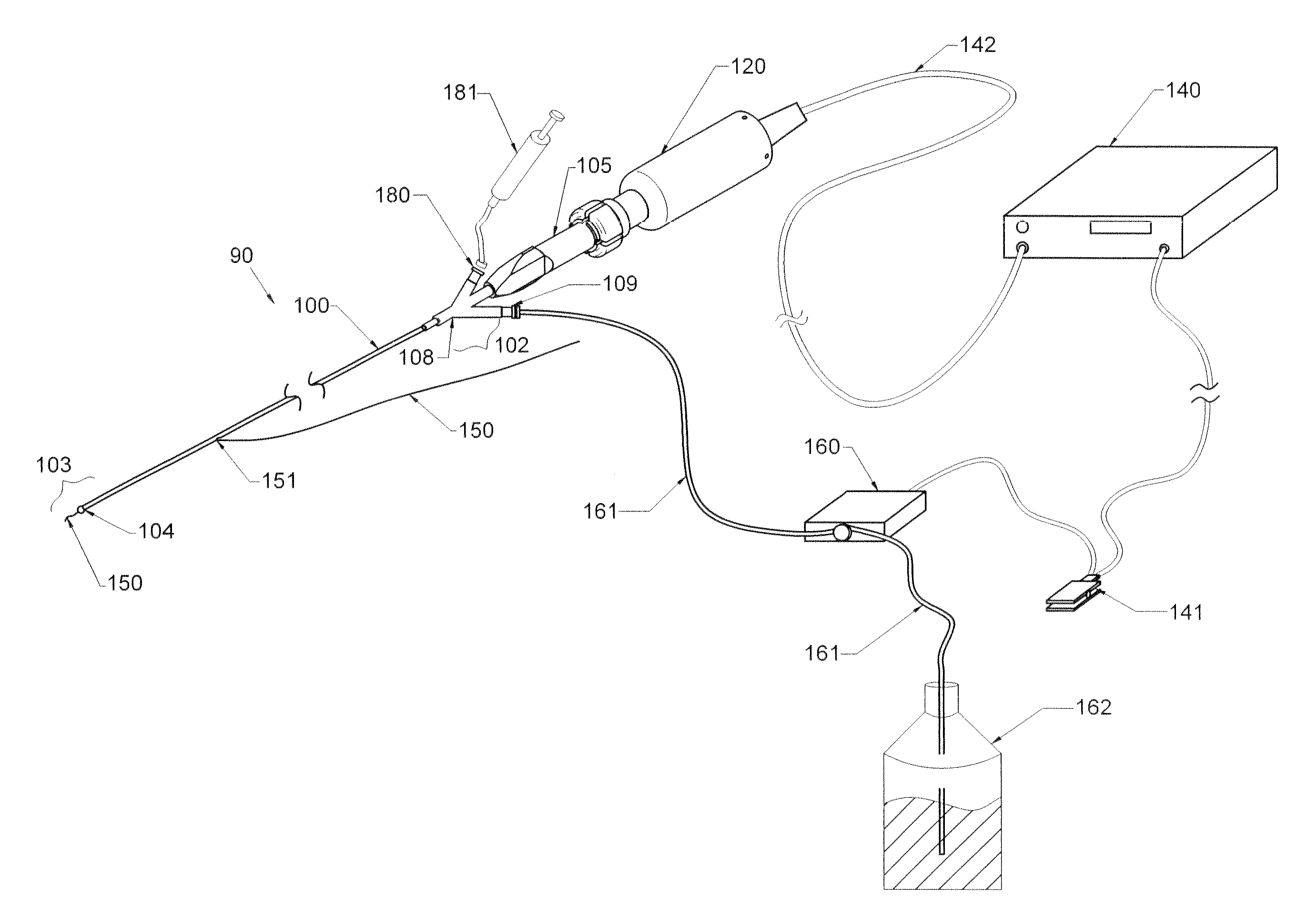
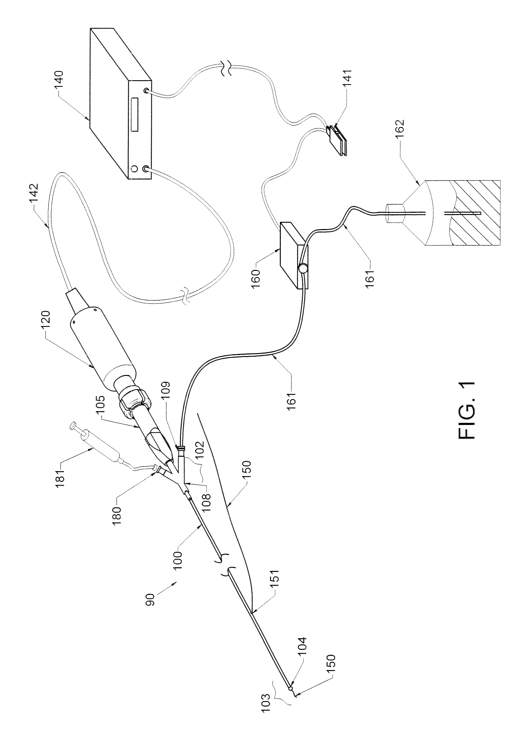
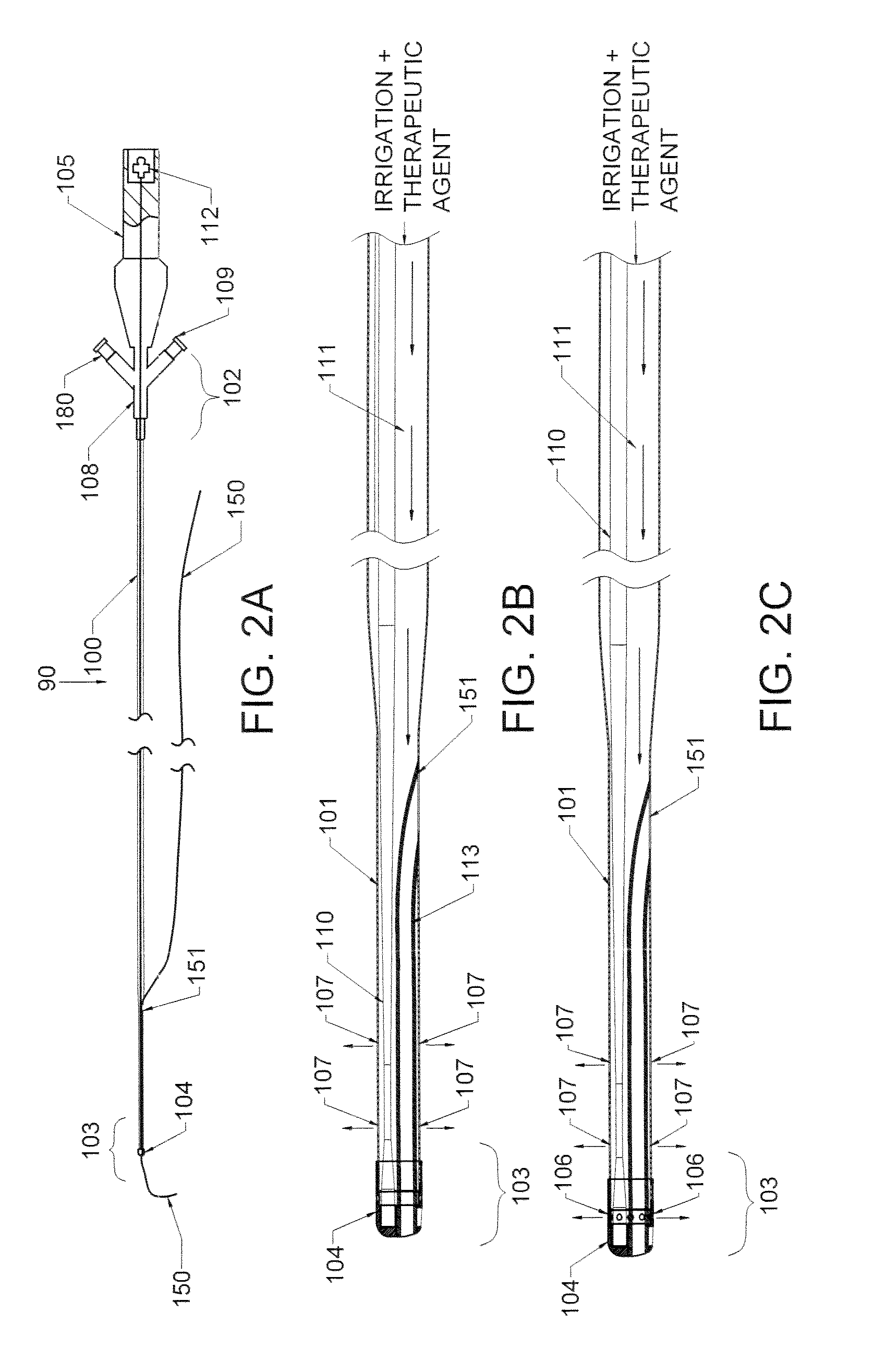
[0081]The present application provides new methods and devices to improve the treatment of vascular stenosis and re-stenosis using ultrasound technology to enhance delivery of therapeutic agents directly to a targeted therapeutic site, such as a stenotic site on an artery or vein wall. These methods may be understood as forms of anti-stenosis treatment, which may include treatment of a stenotic site to modify plaque compliance and to increase the patency of the afflicted vessel, or it may also include treatment of a site previously treated or contemporaneously treated to inhibit or prevent restenosis. Aspects of the invention, including devices and the types of therapeutic agents whose efficacy may be enhanced by the provided technology will be described first in general terms, and then, further below, will be described in the context of FIGS. 1-16.

[0082]The methods described herein employ endovascular sonophoresis and induce vasodilatation, a process that creates micro-indentations...


PUM


Abstract

The present invention provides methods and devices for treating endovascular disease. Vibrational energy is delivered to change compliance and increase permeability at the treatment area. To improve clinical outcomes, one or more therapeutic drugs may be delivered to the treatment area.

Description

CROSS-REFERENCES TO RELATED APPLICATIONS[0001]This application claims priority to US Provisional Application No. 61 / 278,353, of Wallace, filed on Oct. 6, 2009, and is a continuation-in-part of co-pending application Ser. No. 13 / 962,646 filled on Aug. 8, 2013, which is a continuation-in-part of co-pending application Ser. No. 13 / 625,405 filled on Sep. 24, 2012, which is a continuation-in-part of co-pending application Ser. No. 13 / 438,221 filled on Apr. 3, 2012, which is a continuation-in-part of co-pending application Ser. No. 13 / 134,470 filled on Jun. 8, 2011, which is a continuation-in-part of co-pending application Ser. No. 12 / 930,415 filed Jan. 6, 2011, which is a continuation-in-part of co-pending application Ser. No. 12 / 925,495 filed Oct. 22, 2010, which is a continuation-in-part of co-pending application Ser. No. 12 / 807,129, filed Aug. 27, 2010, which is in turn continuation-in-part of co-pending application Ser. No. 12 / 661,853, filed Mar. 25, 2010.TECHNICAL FIELD OF THE INVEN...

Claims


Application Information

Patent Timeline
Patent Type & Authority Applications(United States)
IPC IPC(8): A61B17/22A61M37/00
CPCA61B17/2202A61B17/22004A61B17/22012A61B18/245A61B2017/22014A61B2017/22015A61B2017/22018A61B2017/22039A61B2017/22082A61B2017/22088A61B2017/22098A61B2017/320088A61B2018/00029A61B2018/0212A61M37/0092
Inventor WALLACE, MICHAEL P.
Owner CARDIOPROLIFIC INC
Why Eureka
Try Eureka