

Methods and compositions for typing molecular subgroups of medulloblastoma
a molecular subgroup and typing technology, applied in the field of brain tumor diagnosis, can solve the problems of significant long-term cognitive and/or neuroendocrine adverse effects of survivors, inability to completely remove, and significant cost, and achieve the effect of rapid and accurate identification
- Summary
- Abstract
- Description
- Claims
- Application Information
AI Technical Summary
Benefits of technology
Problems solved by technology
Method used
Image

Examples
example 1
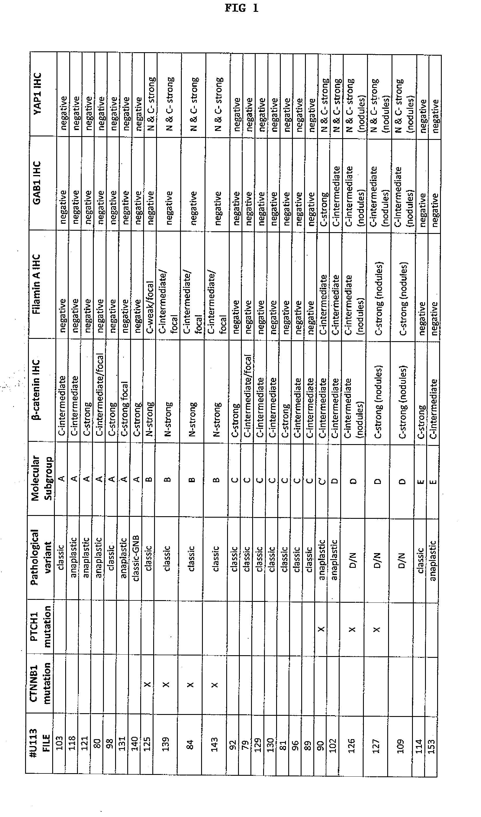
Study Tumor Cohorts
[0140]The main study materials consisted of FFPE tissues from 235 medulloblastomas, representing primary surgical resections from children treated on the SIOP / UKCCSG CNS9102 (PNET3) and CNS9204 trials and from other infants (aged 16 years) treated at Washington University, St. Louis, the Children's Hospital of Los Angeles, and Emory University, Atlanta. The cohort's demographics match those of other previously reported medulloblastoma patient populations. (See Ellison D W, Dalton J, Kocak M, et al: Medulloblastoma: clinicopathological correlates of SHH, WNT, and non-SHH / WNT molecular subgroups. Acta Neuropathol 121:381-96, 2011; herein incorporated by reference in its entirety).
[0141]A separate series of pediatric medulloblastomas (n=26) was used to validate the immunohistochemical assay, the total being limited by availability of FFPE tissue. Gene expression data (Affymetrix U133Av2) were available for the cohort (n=46) from which this validation set was derived....
example 2
Histology and Immunohistochemistry
[0142]Standard histological preparations (hematoxylin & eosin) were used to assess general architectural and cytological features, including nodule formation, differentiation along neuronal (neurocytic / ganglionic) and astrocytic lines, and large cell or anaplastic phenotypes.
[0143]Reticulin preparations were used to evaluate desmoplasia. Internodular desmoplasia was required for a diagnosis of D / N medulloblastoma, including the paucinodular D / N variant, and MBEN. The paucinodular D / N medulloblastoma displays scattered small nodules amid widespread desmoplasia. Intranodular cells in this variant uncommonly demonstrate the differentiated neurocytic phenotype of the conventional D / N tumor, but do express neuronal proteins and show low Ki-67 immunolabeling. The MBEN is defined by its large irregularly shaped nodules, pronounced internodular neurocytic differentiation, and sparse internodular desmoplastic regions.
[0144]As defined by the WHO classificatio...
example 3
Frequencies and Phenotypes Among Pathological Variants—Defining the Tumor Cohort
[0147]Classic medulloblastomas dominated the study cohort, accounting for 72% of all tumors. Most classic tumors (86%) appeared as sheets of uniform small cells with a high nuclear:cytoplasmic ratio and round hyperchromatic nuclei, while the remainder (14%) had a dominant spindle-cell morphology. Focal neuronal differentiation was evident in some classic tumors, manifesting either as nodules of uniform neurocytic cells without surrounding reticulin-positive desmoplasia (non-desmoplastic nodular ‘biphasic’ phenotype; 7%), or as dense clusters of tiny round cells (4%), or as foci of neuropil-like matrix with an irregular border, variable area, and scattered ganglion or neurocytic cells (ganglioneuroblastoma phenotype; n=1). In these tumors, foci of neuronal differentiation demonstrated: (i) the expected moderate to strong immunoreactivities for synaptophysin and NEU-N, (ii) up-regulation of p2′7, and (iii)...
PUM
| Property | Measurement | Unit |
|---|---|---|
| size | aaaaa | aaaaa |
| frequency | aaaaa | aaaaa |
| morphology | aaaaa | aaaaa |
Abstract
Description
Claims
Application Information