Imaging system

Active Publication Date: 2014-07-03
IND TECH RES INST
View PDF3 Cites 11 Cited by
  • Summary
  • Abstract
  • Description
  • Claims
  • Application Information

AI Technical Summary

Benefits of technology

[0008]In light of the above, in the imaging system described in the embodiment, the X-ray source which provides a multienergy polychromatic X-ray is disposed and the multienergy bands of the sensor correspond to the multienergy bands of the X-ray. In such configuration, through phase contrast, the imaging system can ac

Problems solved by technology

However, because the composition differences of soft tissues at various parts of the body are not significant, the image differences within the X-ray energy range of bone radiography caused by the composition differences between the soft tissues are not significant as well.
As a result, the soft tissue imaging result is not easily distinguished and is difficult to serve for medical diagn

Method used


Image

  • Imaging system
  • Imaging system
  • Imaging system

Examples


Example

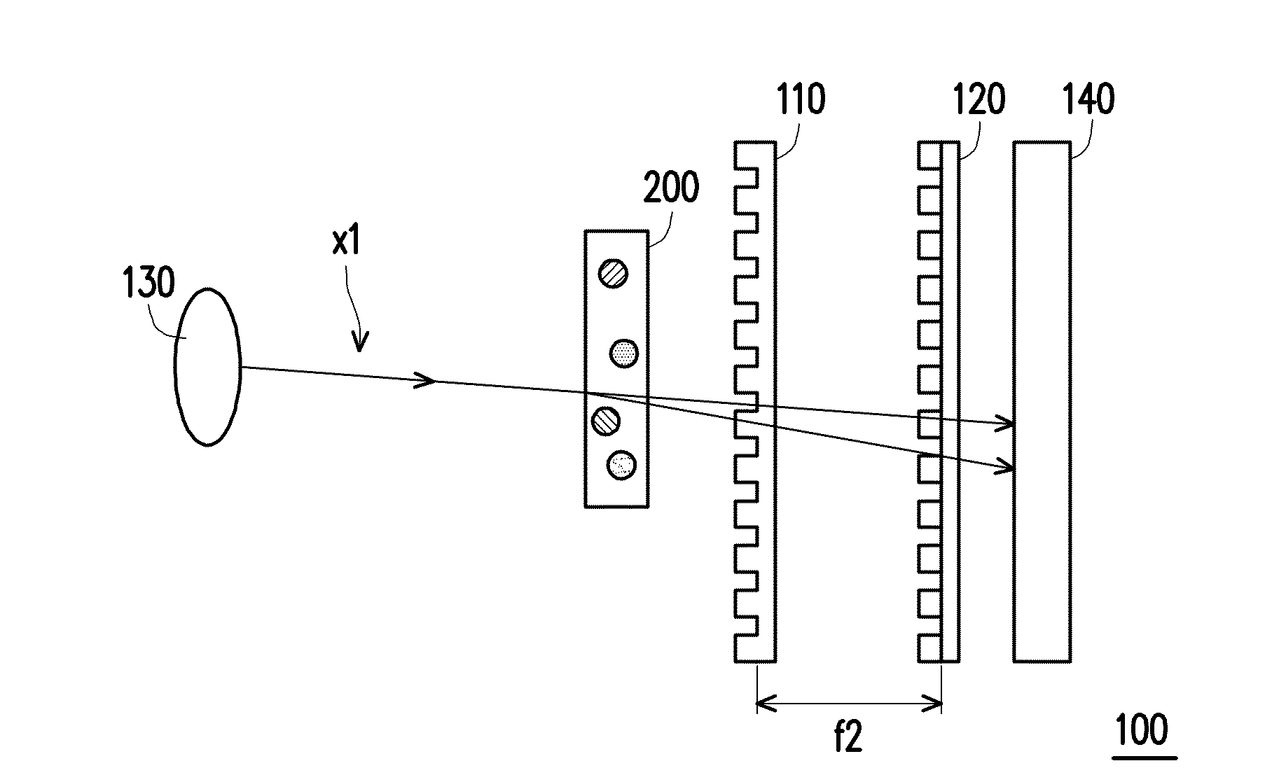
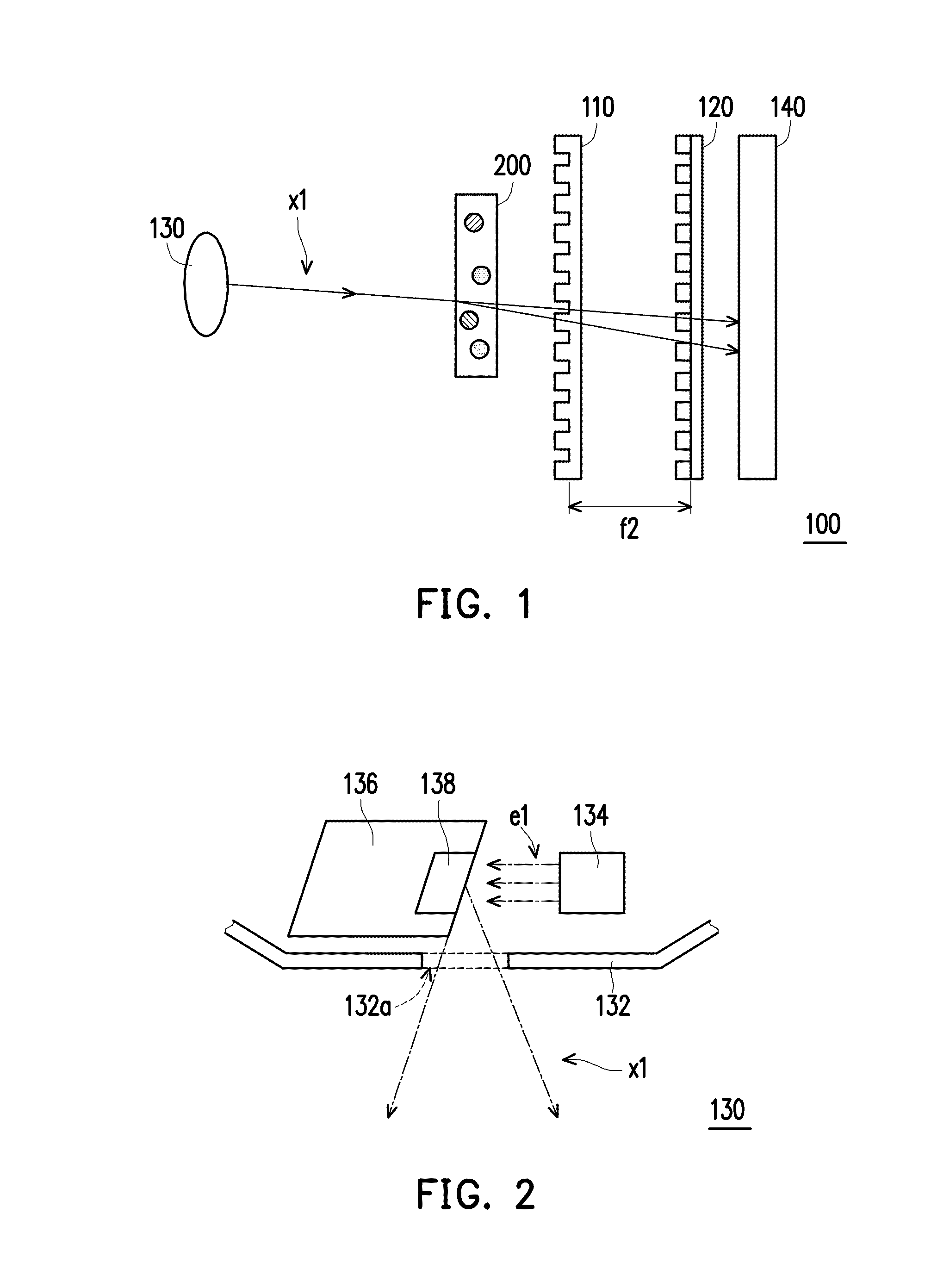
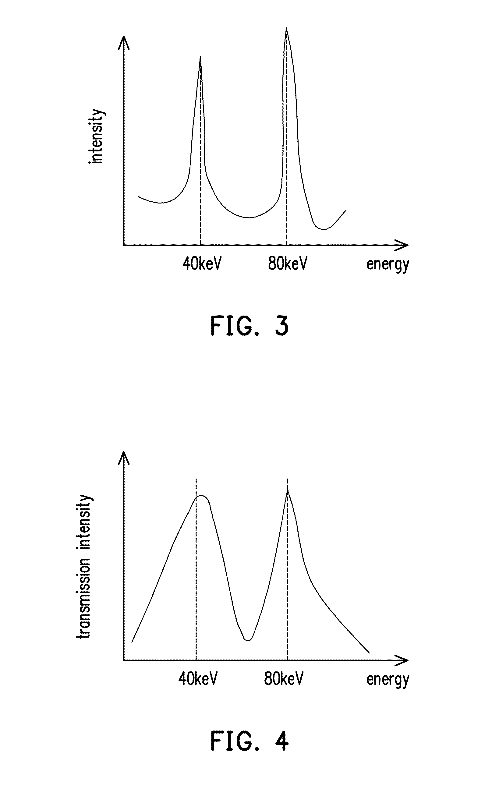
[0023]FIG. 1 is a schematic view of an X-ray imaging system according to an exemplary embodiment of the disclosure. FIG. 2 is a schematic view of the X-ray source of the imaging system of FIG. 1, and only parts of components are shown for equivalent representing. FIG. 3 is the X-ray spectrogram at the X-ray source of the X-ray imaging system of FIG. 1. FIG. 4 is the X-ray spectrogram detected by the sensor of the X-ray imaging system of FIG. 1. Referring to FIG. 1 through FIG. 4 together, in the embodiment, the imaging system 100 includes an X-ray source 130, a first light grating 110, a second light grating 120 and a sensor 140, wherein an object 200 is adapted to be disposed between the X-ray source 130 and the sensor 140, the first light grating 110 is disposed between the object 200 and the sensor 140, and the second light grating 120 is disposed between the first light grating 110 and the sensor 140. The X-ray source 130 is a characteristic radiation and used for generating mul...


PUM


Abstract

An imaging system used for material discrimination of an objet is provided. The imaging system includes an X-ray source and an imaging sensor. The X-ray source generates multienergy polychromatic X-ray, wherein there is an integral-multiple relationship between the multi-energy-band of the X-ray. The object is placed between the X-ray source and the imaging sensor. The X-ray is transmitted toward and imaged by the imaging sensor through the object. An imaging multi-energy-band of the imaging sensor corresponds to the multi-energy-band of the X-ray.

Description

CROSS-REFERENCE TO RELATED APPLICATION[0001]This application claims the priority benefits of U.S. provisional application Ser. No. 61 / 739,017, filed on Dec. 19, 2012 and Taiwan application serial no. 102142336, filed on Nov. 20, 2013. The entirety of each of the above-mentioned patent applications is hereby incorporated by reference herein and made a part of this specification.FIELD OF THE DISCLOSURE[0002]The disclosure relates to an imaging system, and particularly relates to an X-ray phase contrast imaging system.DESCRIPTION OF RELATED ART[0003]X-ray (i.e., X-light) medical imaging is a non-invasive method for checking the structure inside a human body and it can be quickly aware of the anatomy information of a subject (such as shape structures of bones, organs and soft tissues) without performing anatomy practice or tissue sectioning. Therefore, X-ray medical imaging result has served as one of the bases of medical diagnosis.[0004]The conventional X-ray imaging technology uses X-...

Claims


Application Information

Patent Timeline
IPC IPC(8): G01N23/04
CPCG01N23/04G01N23/087A61B6/4035A61B6/4241A61B6/482A61B6/484G01N2223/423
Inventor LU, HUI-HSINGUO, JIUN-LINWANG, WEI-HSINCHEN, TING-HSUANKUO, TSUNG-TER
Owner IND TECH RES INST
Why Eureka
Try Eureka