System and method for the classification of healthiness index from chest radiographs of a healthy person

Inactive Publication Date: 2017-11-23
SHENZHEN SMART IMAGING HEALTHCARE
View PDF3 Cites 5 Cited by
  • Summary
  • Abstract
  • Description
  • Claims
  • Application Information

AI Technical Summary

Benefits of technology

[0008]An embodiment of the present disclosure discloses a method for classifying healthiness indices in a normal radiological image. The method comprises the steps of: image pre-processing comprising image enhancement and normalization processing to enhance image contrast; image segmentation to identify body parts and boundaries thereof, the image segmentation step further comprising a sub-step of differentiation of different

Problems solved by technology

A chest X-ray may not be able to

Method used


Image

  • System and method for the classification of healthiness index from chest radiographs of a healthy person
  • System and method for the classification of healthiness index from chest radiographs of a healthy person
  • System and method for the classification of healthiness index from chest radiographs of a healthy person

Examples


Example

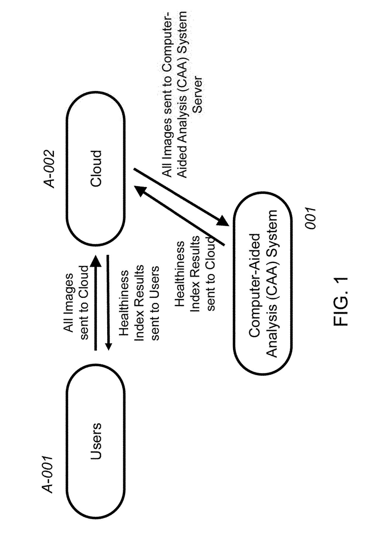
[0028]FIG. 1 illustrates a schematic block diagram of a medical non-diagnostic or diagnostic workflow environment incorporating a computer-aided-analysis CAA) system 001 between users A-001 and internet cloud A-002.

[0029]The users A-001 sends all images to the cloud from the image acquisition systems, computers, smartphone, media, storage device, to internet cloud A-002. The interne cloud A-002 sends all images to Computer-Aided Analysis (CAA) System Server. The CAA system performs the classification of healthiness index and sends the classification results back to the cloud A-002. The cloud A-002 then sends the healthiness index to the users A-001.

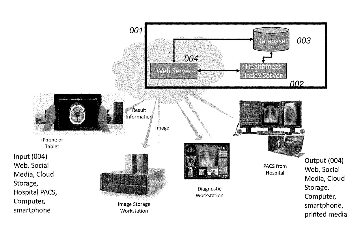
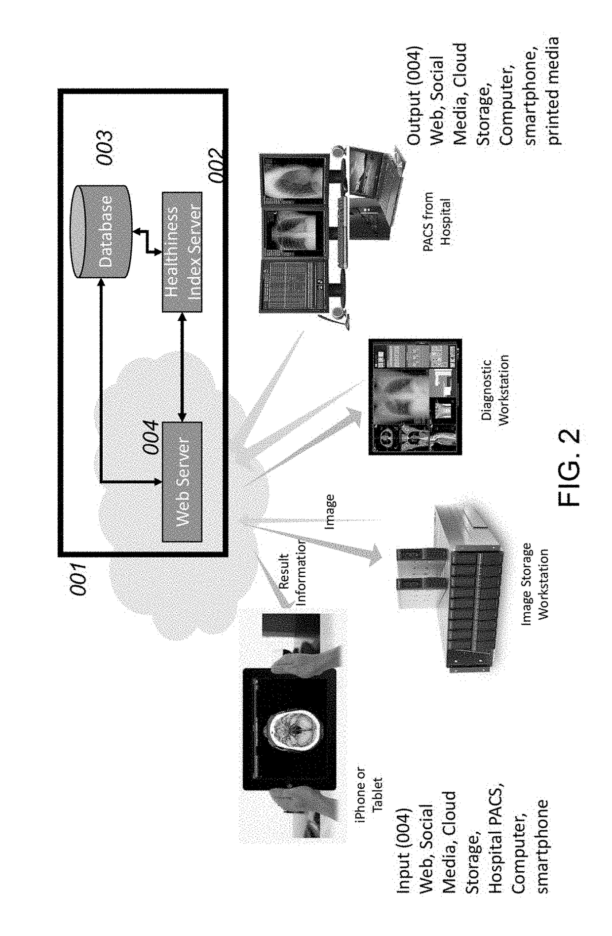
[0030]FIG. 2 illustrates a schematic block diagram of a medical non-diagnostic or diagnostic environment incorporating a computer-aided-analysis (CAA) system between a non-diagnostic or diagnostic medical imaging acquisition system, individual computer system, smartphone, storage device, media and an archive / review static.

[0031]Input 004 ...


PUM


Abstract

A method for classifying the degree of healthiness from chest radiographs comprises inputting image data with a medical imaging acquisition system or from individual's computers or smartphones or cloud storage devices. The image data is transmitted from the medical imaging acquisition system or from the computers or storage device to a computer-aided-analysis (CAA) system via the Internet and an archive/review station. Classification results are generated by processing the image data to perform lung segmentation and generate various radiomics, perform classification of radiomics. The classification results are transmitted from the CAA system to archive/review servers or the computers/smartphones via the Internet. The classification results are used to retrieve the associated clinical, wellness, and health knowledge from the database to form composite data. The composite data is sent to end users including patients, healthcare providers, consultants, and other authorized personnel and displayed by the archive/review system or the computers/smartphones.

Description

FIELD OF THE INVENTION[0001]The present invention relates to an automated method and system for digital image processing of radiologic images and, more specifically, to an automated method and system for the classification of different healthiness indices, using radiomics, digital image processing and artificial intelligence.BACKGROUND OF THE INVENTION[0002]Many companies and government agencies offer annual physical exams to every people, regardless the age, as part of their fringe benefits or prior to their employment. Chest X-ray is sometimes included in the physical exam. Although most of people will be normal on the chest ray, people are very concerning about their potential development of heart and lung diseases especially lung cancer due to high air pollution and would like to prevent or slow down the trend to develop lung cancer. Lately, more and more of people have obtained their digital chest X-ray images to store in their computers, smart phones, or cloud storage devices....

Claims


Application Information

Patent Timeline
IPC IPC(8): G06T7/00G06T7/11G06K9/46G06K9/62G06T7/13
CPCG06T7/0012G06K9/628G06T7/11G06T2200/28G06T7/13G06T2207/10116G06T2207/30061G06K9/46G06T7/12A61B6/50A61B5/0013A61B5/08A61B5/7275A61B6/5217A61B6/5294A61B6/563G16H50/30G06V2201/031A61B5/00G06F18/2431
Inventor LURE, YUAN-MING FLEMINGZHOU, HAO
Owner SHENZHEN SMART IMAGING HEALTHCARE
Why Eureka
Try Eureka