
Apparatus and method for lesion segmentation in medical image

Active Publication Date: 2014-08-28
SEOUL NAT UNIV R&DB FOUND
View PDF7 Cites 26 Cited by
  • Summary
  • Abstract
  • Description
  • Claims
  • Application Information

AI Technical Summary

Benefits of technology

The patent describes a method for identifying and segmenting abnormal areas in medical images. It uses a series of parameters to emphasize slopes, separate out lesion regions from non-lesion regions, and filter out noise. This approach helps to improve the accuracy and efficiency of identifying potential lesions in images.

Problems solved by technology

However, the point-based segmentation method requires a user to manually move control points.

Method used


Image

  • Apparatus and method for lesion segmentation in medical image
  • Apparatus and method for lesion segmentation in medical image
  • Apparatus and method for lesion segmentation in medical image

Examples


Embodiment Construction

[0040]The following description is provided to assist the reader in gaining a comprehensive understanding of the methods, apparatuses, and / or systems described herein. Accordingly, various changes, modifications, and equivalents of the methods, apparatuses, and / or systems described herein will be suggested to those of ordinary skill in the art. Also, descriptions of well-known functions and constructions may be omitted for increased clarity and conciseness.

[0041]The terminology used herein is for the purpose of describing particular embodiments only and is not intended to be limiting of the present invention. As used herein, the singular forms “a,”“an” and “the” are intended to include the plural forms as well, unless the context clearly indicates otherwise. It will be further understood that the terms “comprises” and / or “comprising,” when used in this specification, specify the presence of stated features, integers, steps, operations, elements, and / or components, but do not preclud...


PUM


Abstract

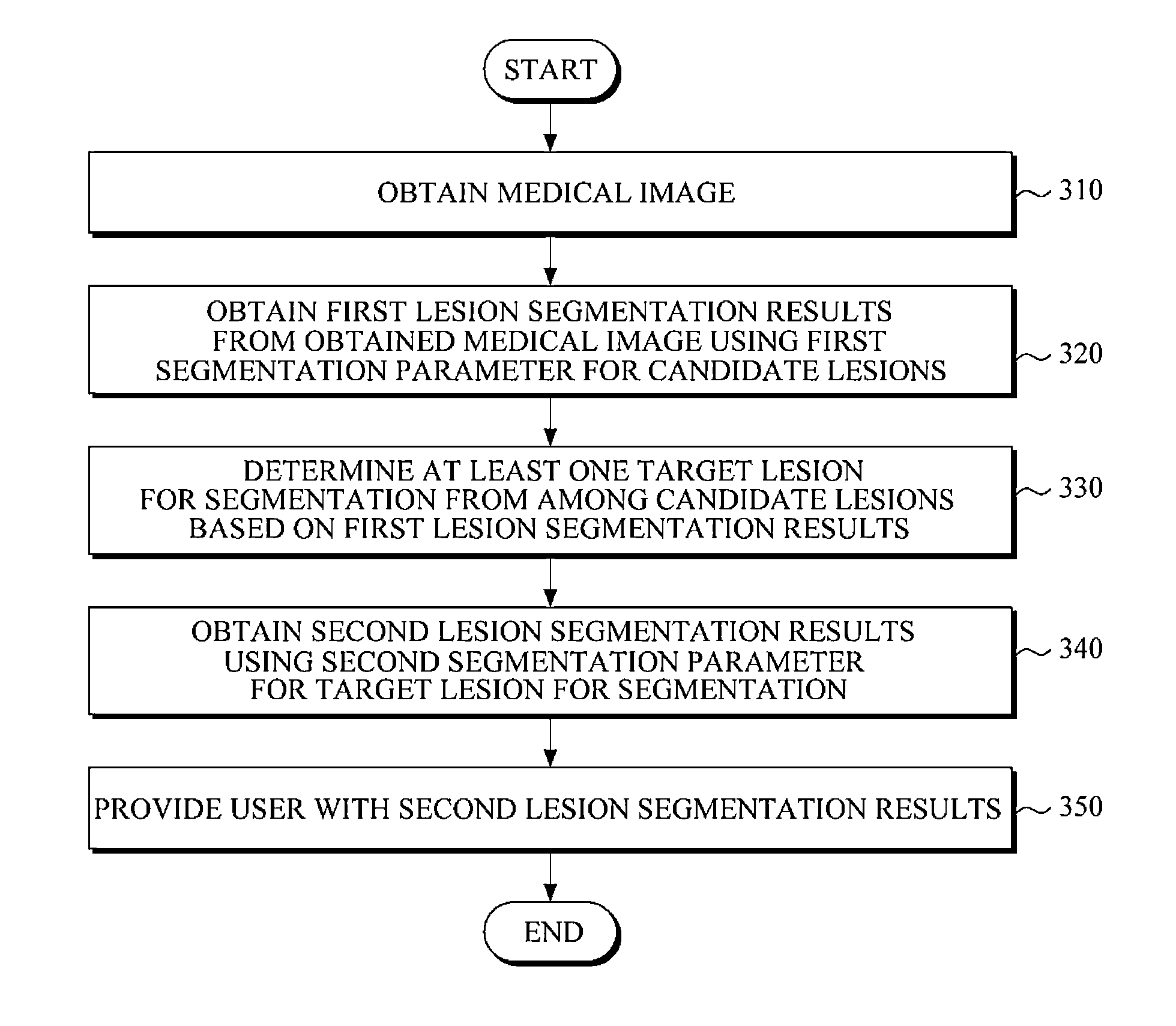
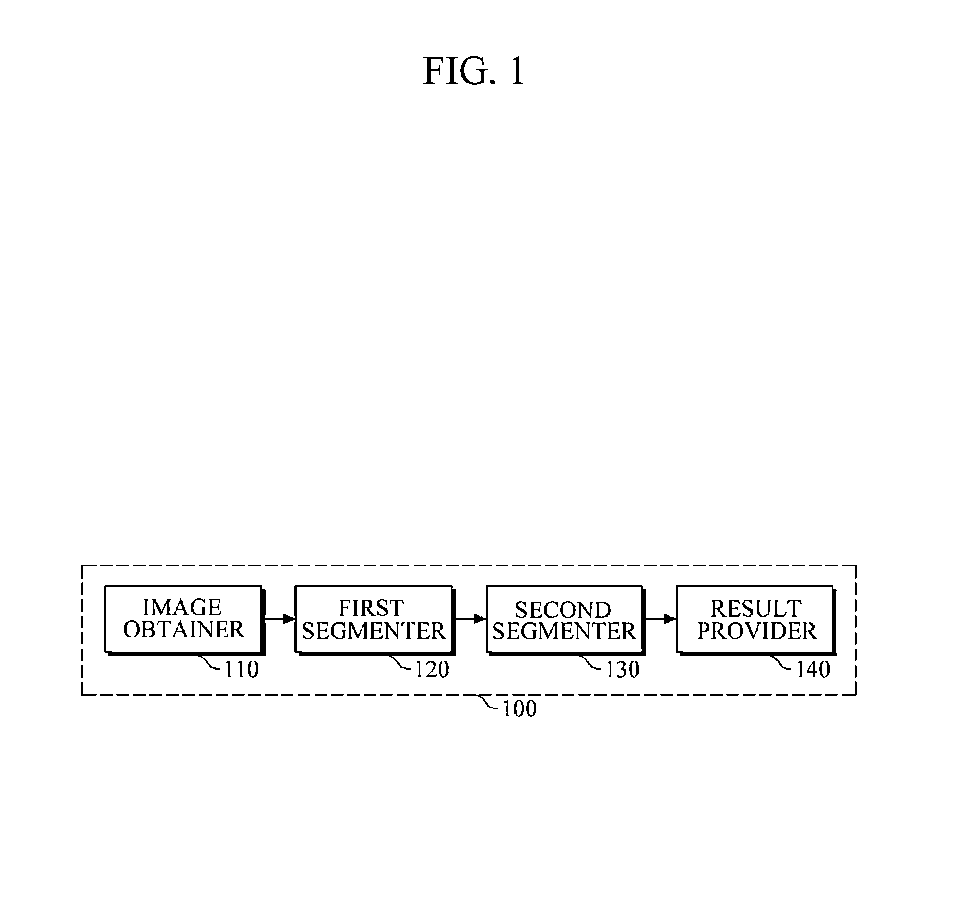
An apparatus and method are provided including a first segmenter and a second segmenter. The first segmenter is configured to generate a first segmentation result from a medical image using a first segmentation parameter for a candidate lesion. The second segmenter is configured to determine a target lesion to segment from among the candidate lesion based on the first segmentation result, and generate a second segmentation result using a second segmentation parameter to segment the target lesion.

Description

CROSS-REFERENCE TO RELATED APPLICATION(S)[0001]This application claims the benefit under 35 U.S.C. §119(a) of a Korean Patent Application No. 10-2013-0019930, filed on Feb. 25, 2013, in the Korean Intellectual Property Office, the entire disclosure of which is incorporated herein by references for all purposes.BACKGROUND[0002]1. Field[0003]The following description relates to an apparatus and method to segment a lesion in a medical image.[0004]2. Description of Related Art[0005]A Computer Aided Diagnosis (CAD) system is used to detect and diagnose a lesion from an enormous amount of medical images. The CAD system segments a medical image including a region suspected to include a lesion, and provides a user with information about the lesion region and a location thereof. If the lesion segmentation information provided by the CAD system does not conform to criterion discrimination defined by the user, the user ignores the lesion segmentation, and manually segments again the region sus...

Claims


Application Information

Patent Timeline
IPC IPC(8): G06T7/00
CPCG06T7/0081G06T2207/30096G06T7/11A61B6/00G06T7/00
Inventor PARK, MOON-HOLEE, MYUNG-EUNKIM, YE-HOONKIM, JONG-HYOKIM, JUNG-HOESEONG, YEONG-KYEONGYU, SIN-SANGCHO, BAEK-HWANJIN, HYEONG-MIN
Owner SEOUL NAT UNIV R&DB FOUND
Learn More