High-throughput single-cell imaging, sorting, and isolation

a single-cell, high-throughput technology, applied in the field of microfluidic methods and apparatuses, can solve the problems of difficult automation, slow gradient methods, and difficult characterization and isolation of individual target cells

Active Publication Date: 2014-09-04
CAPLIPER LIFE SCI INC
View PDF5 Cites 15 Cited by
  • Summary
  • Abstract
  • Description
  • Claims
  • Application Information

AI Technical Summary

Benefits of technology

[0009]One aspect of the present invention is an apparatus for isolating individual target cells. The apparatus comprises a body structure comprising a main channel, a collection channel, and a waste channel fluidly coupled at a first fluid junction. A plurality of trapping channels intersect the collection channel, each of the trapping channels having a diameter at a location adjacent to the intersection of the trapping channel with the collection channel that is less than a diameter of an individual targe...

Problems solved by technology

Unfortunately, both WBCs and CTCs are rare in whole-blood samples, making their characterization and isolation problematic.
While effective, the gradient methods are typically slow, difficul...

Method used


Image

  • High-throughput single-cell imaging, sorting, and isolation
  • High-throughput single-cell imaging, sorting, and isolation
  • High-throughput single-cell imaging, sorting, and isolation

Examples


Embodiment Construction

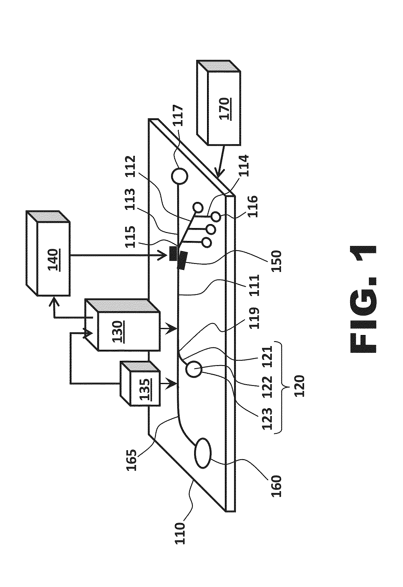
[0015]One aspect of the present invention is an apparatus for isolating individual target cells. One apparatus in accordance with the present invention is seen in FIG. 1 as comprising a body structure 110, a cell focusing system 120, an imaging system 130, a lighting and / or stimulating source 135 that operates in association with the imaging system, a processor 140, a directing system 150, a cell source 160, a sample enrichment system 165, and a pressure source 170.

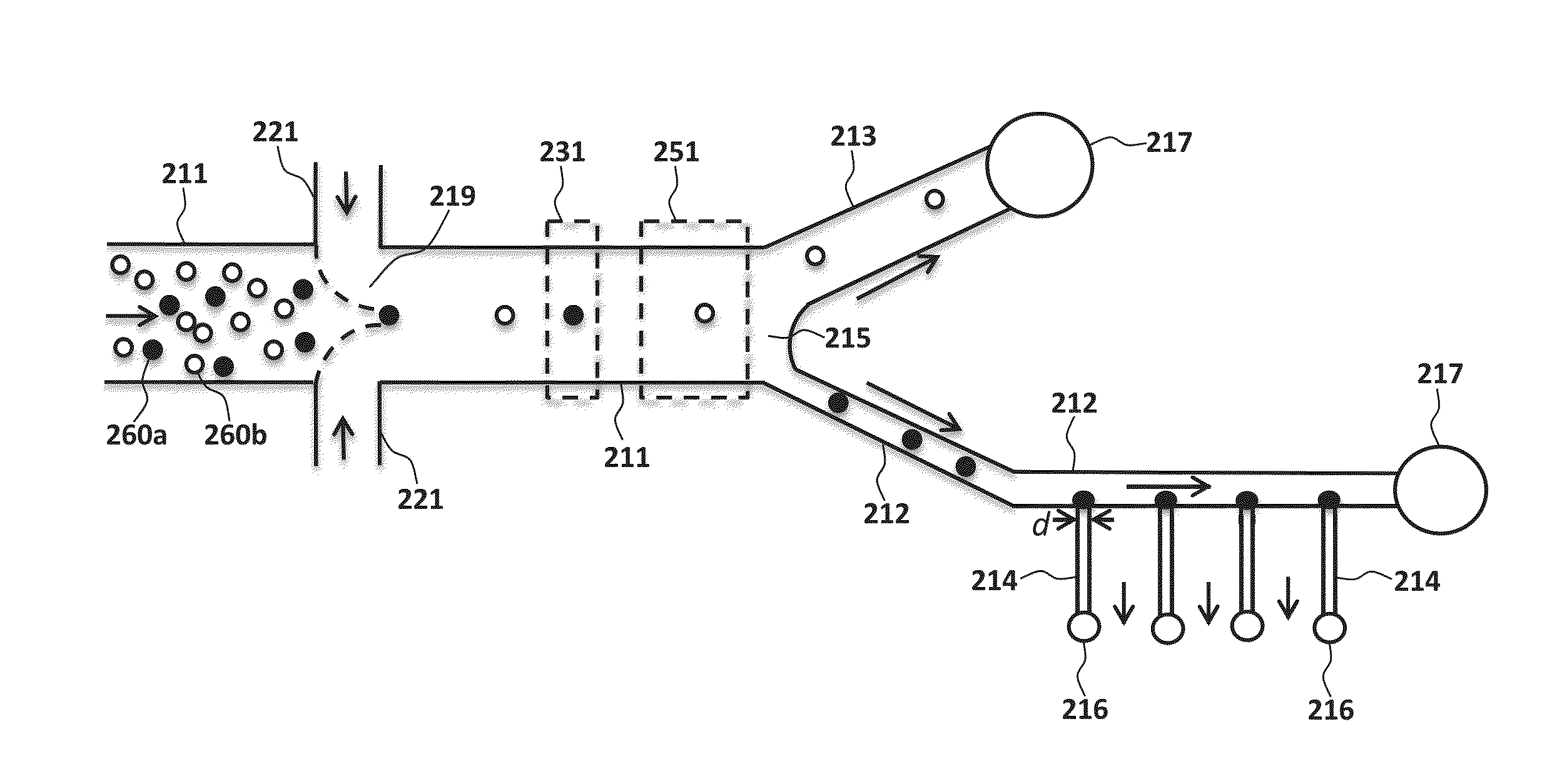
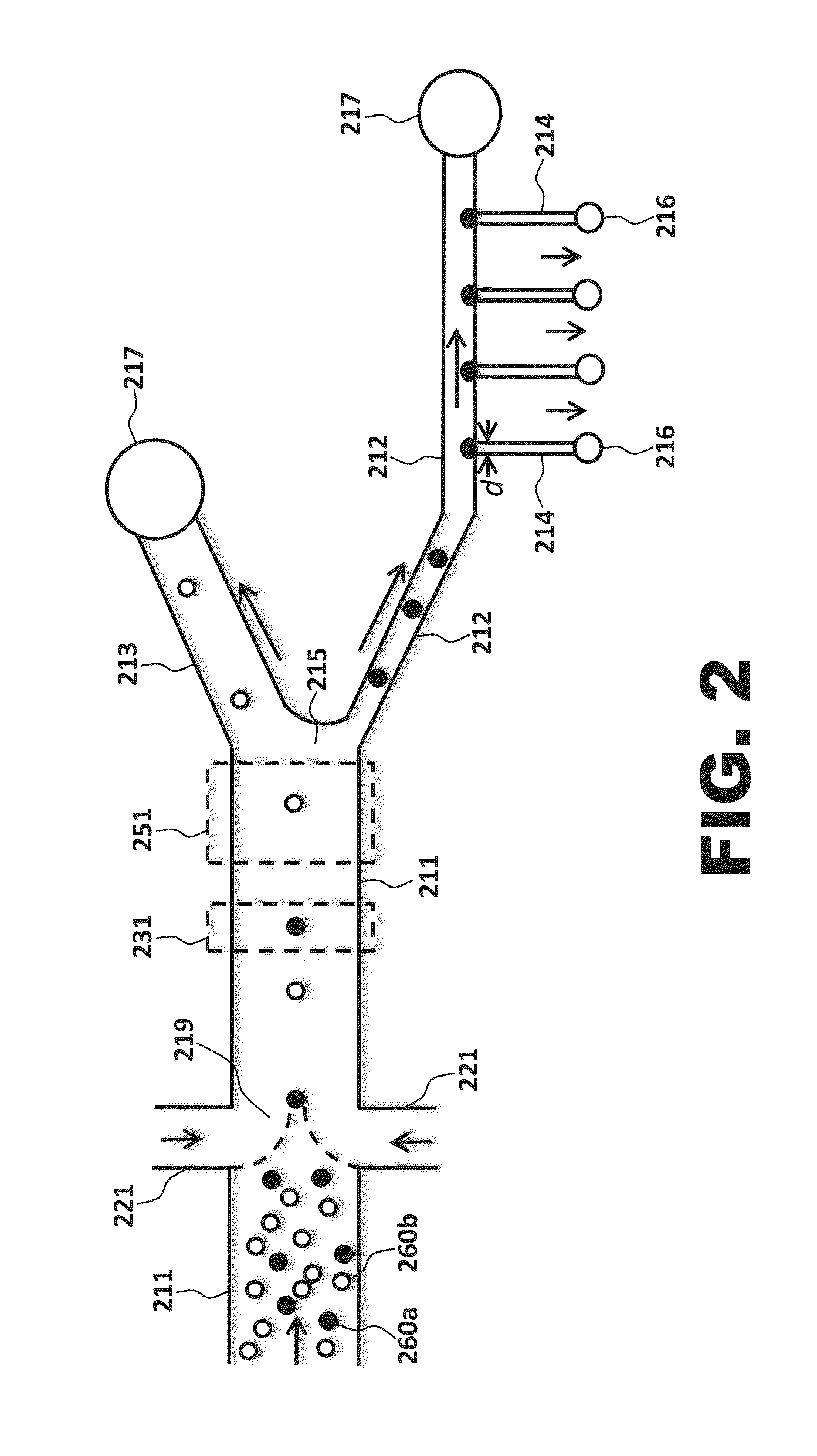
[0016]FIGS. 1 and 2 both illustrate body structures according to the present invention. While the outer edges of the body structure are not shown in FIG. 2, the elements illustrated in the figure are to be understood as being disposed in or on a body structure. Either or both of the illustrated body structures may be microfluidic; i.e., the body structure may be engineered to operate using small volumes of fluid (typically μ1 volumes of fluid) and / or may have at least one channel with a diameter ≦1 mm.

[0017]As seen in FIG...


PUM


Abstract

The invention provides a method and apparatus for isolating individual target cells. The apparatus includes a body structure comprising a main channel, a collection channel, and a waste channel fluidly coupled at a first fluid junction. A plurality of trapping channels intersect the collection channel, each trapping channel having a diameter at a location adjacent to the intersection of the trapping channel with the collection channel that is less than a diameter of an individual target cell. The apparatus also includes an imaging system configured to image individual target and non-target cells within the main channel, thereby producing imaging data; a processor configured to perform real-time, multivariate analyses of the imaging data; and a directing system configured to direct the individual target cells. A pressure source is in fluid communication with one or more of the collection channel, the waste channel, the first side channel, and the second side channel.

Description

TECHNICAL FIELD[0001]The present disclosure is in the field of microfluidic methods and apparatuses. In particular, described herein are methods and apparatuses for imaging, sorting, and isolating individual target cells.BACKGROUND OF THE INVENTION[0002]High-content analysis and isolation of cells is a growth area in personalized medicine. Both white blood cells (WBCs) and circulating tumor cells (CTCs), for example, can provide valuable information for diagnosis and treatment of diseases.[0003]High throughput screening of WBCs can help determine whether a sick patient is responding to a specific drug or a healthy individual has mounted an adequate response to an immunization. Isolated viable WBCs can be used to determine whether specific T cell subpopulations are present in the blood and are capable of eliciting an immune response to the human immunodeficiency virus (HIV).[0004]CTCs, i.e., tumor cells that are identified in transit within the blood stream, are shed from primary and...

Claims


Application Information

Patent Timeline
IPC IPC(8): G06T7/00
CPCG06T7/0012B01L3/502761C12M47/04B01L2200/0636B01L2200/0652B01L2200/0668B01L2300/0816B01L2300/0864C12N5/0081H04N23/56C12N5/0087H04N7/18G02B21/365
Inventor BHARADWAJ, RAJIVFATHOLLAHI, BAHRAM
Owner CAPLIPER LIFE SCI INC
Why Eureka
Try Eureka