Image quality assessment of microscopy images

Active Publication Date: 2014-09-18
LEICA MICROSYSTEMS CMS GMBH
View PDF5 Cites 12 Cited by
  • Summary
  • Abstract
  • Description
  • Claims
  • Application Information

AI Technical Summary

Benefits of technology

[0006]In one embodiment, a computer-implemented method for assessing image quality is provided. The method includes the act of acquiring a first image and a second image. At least a portion of the first image and the second image overlap. A rotation and a scale are determined relating the first image and the second image. A respective Fourier transform of the first imag

Problems solved by technology

However, the capability of acquiring imagery for a large number of biomarkers results in a large number of images being acquired.
As will be appreciated, so

Method used


Image

  • Image quality assessment of microscopy images
  • Image quality assessment of microscopy images
  • Image quality assessment of microscopy images

Examples


Embodiment Construction

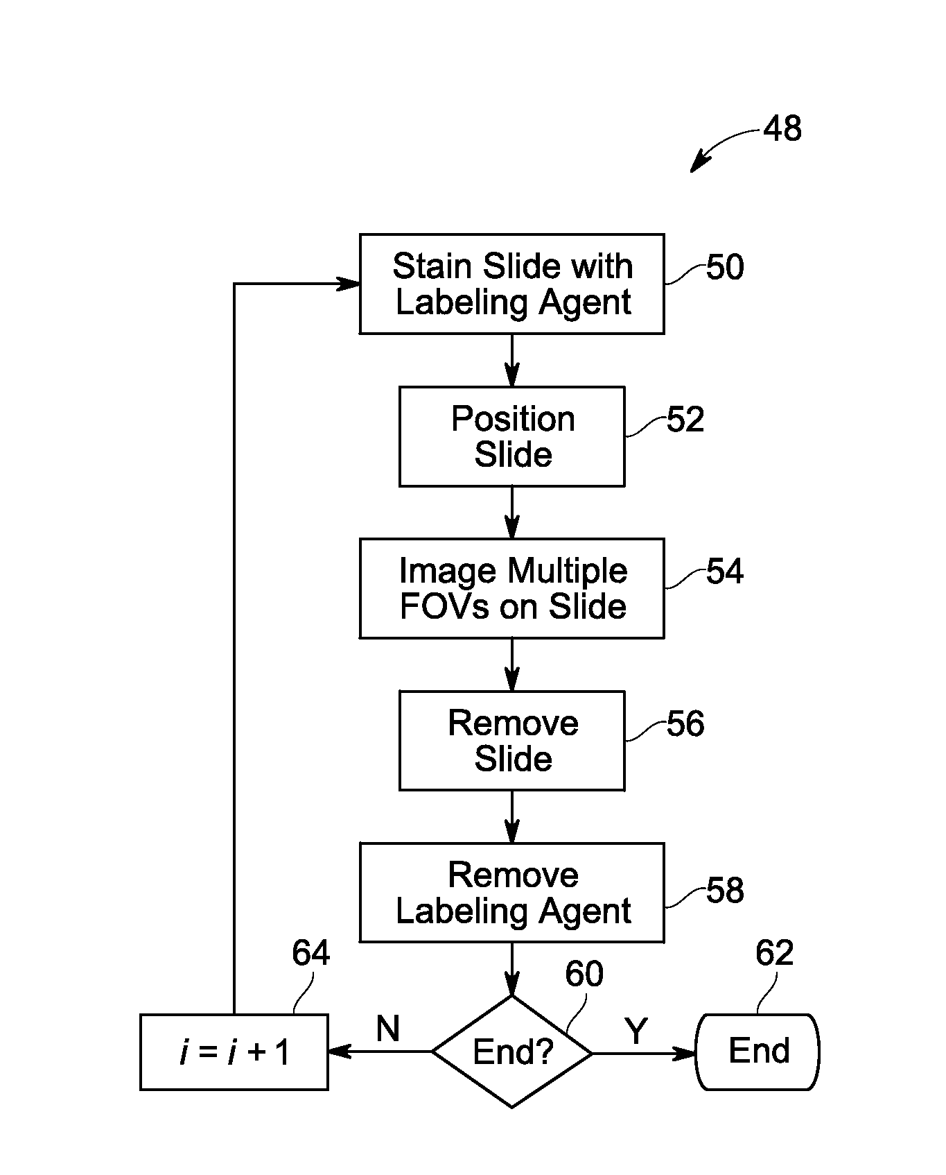
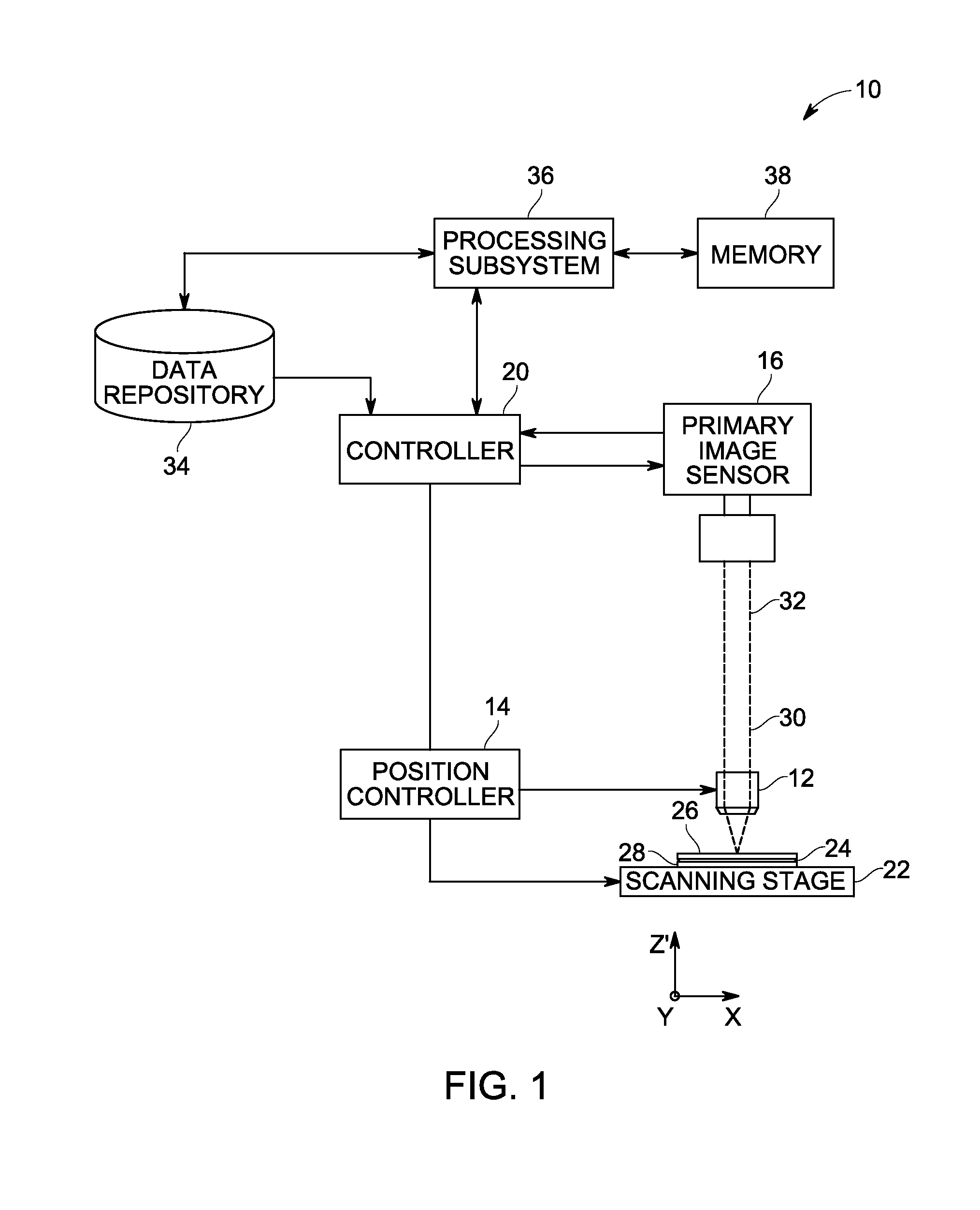
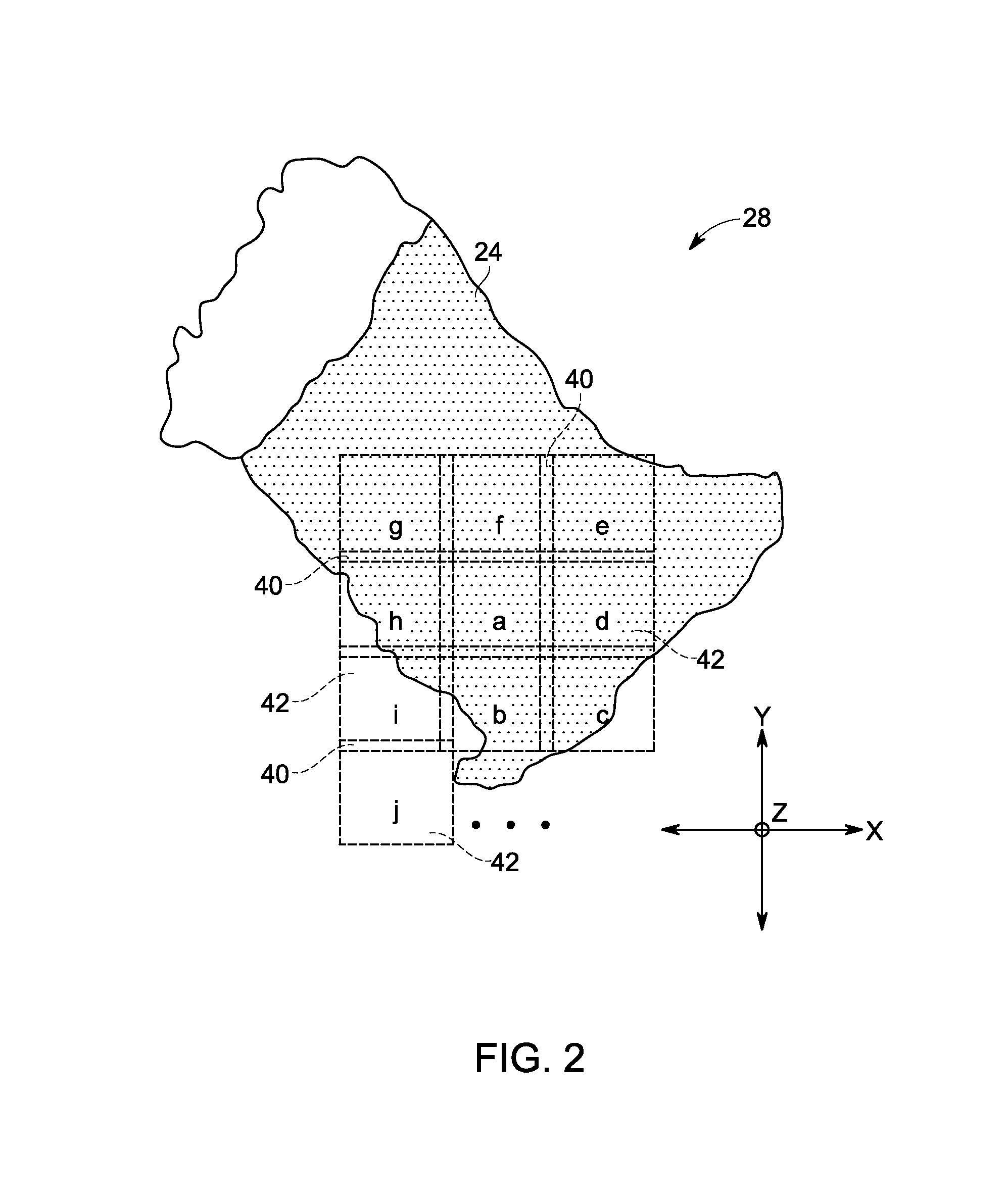
[0018]The large number of images produced by automated, multiplexed scanning devices (such as may be used in immunohistochemical studies) makes manual detection of imaging failures—both gross failures of focus and position, and partial-image artifacts such as damaged tissue and foreign objects—difficult, if not infeasible. As such, it may be desirable to automate the detection of imaging failures. With this in mind, the present approach describes a receiver pipeline that, in one embodiment, registers images using rigid-body transformations in the Fourier domain, detects whole-image defects based on the figure of merit from the registration operation, and detects partial-image defects by calculating correlation in local regions of the image. As discussed herein, in accordance with the present approach, the most common problems with the images can be identified by automatic examination. Defective images (or parts of images) can then be excluded from statistical analysis to avoid conta...


PUM


Abstract

Automated assessment of registration quality, focus, and area defects in sequentially acquired images, such as images acquired by a digital microscope, is disclosed. In one embodiment, acquired images are registered and whole-image defects are automatically detected based on a figure of merit generated by the registration process. In related implementations, area defects may be automatically detected by calculating correlations in localized image regions for images acquired in different imaging rounds.

Description

BACKGROUND[0001]The subject matter disclosed herein relates to the assessing the quality of microscopy images.[0002]For various physiological conditions, such as cancer, infectious diseases, physiological disorders, and so forth, detection and monitoring may be based, in part, on the analysis of a biological specimen from the patient. For example, a sample may be analyzed to detect the presence of abnormal numbers or types of cells and / or organisms that may be indicative of a disease or disorder. Various types of microscopy may be employed for such analysis. Further, various stains and staining protocols may be employed as part of this analysis to allow visualization of different structures, chemicals, or environments that might aid in detection or diagnosis of a disease or disorder.[0003]To facilitate analysis of such pathology or histology samples, automated microscopy systems have been developed that automate various aspects of the image acquisition process. In particular, digita...

Claims


Application Information

Patent Timeline
IPC IPC(8): G06T7/00
CPCG06T7/0012G06T2200/32G06T2207/30024G06T2207/30072G06T7/37G06T7/0002G06V20/693G06V10/993
Inventor KENNY, KEVIN BERNARDROTHNEY, MEGAN PEARL
Owner LEICA MICROSYSTEMS CMS GMBH
Why Eureka
Try Eureka