Ophthalmologic imaging apparatus, method of controlling the same, and program
a technology of ophthalmologic imaging and control method, applied in the field of ophthalmologic imaging apparatus, can solve the problems of increased apparatus size, lack of flexibility of technique, high cost, etc., and achieve the effect of favorable manual focus operability
- Summary
- Abstract
- Description
- Claims
- Application Information
AI Technical Summary
Benefits of technology
Problems solved by technology
Method used
Image
Examples
first embodiment
[0025
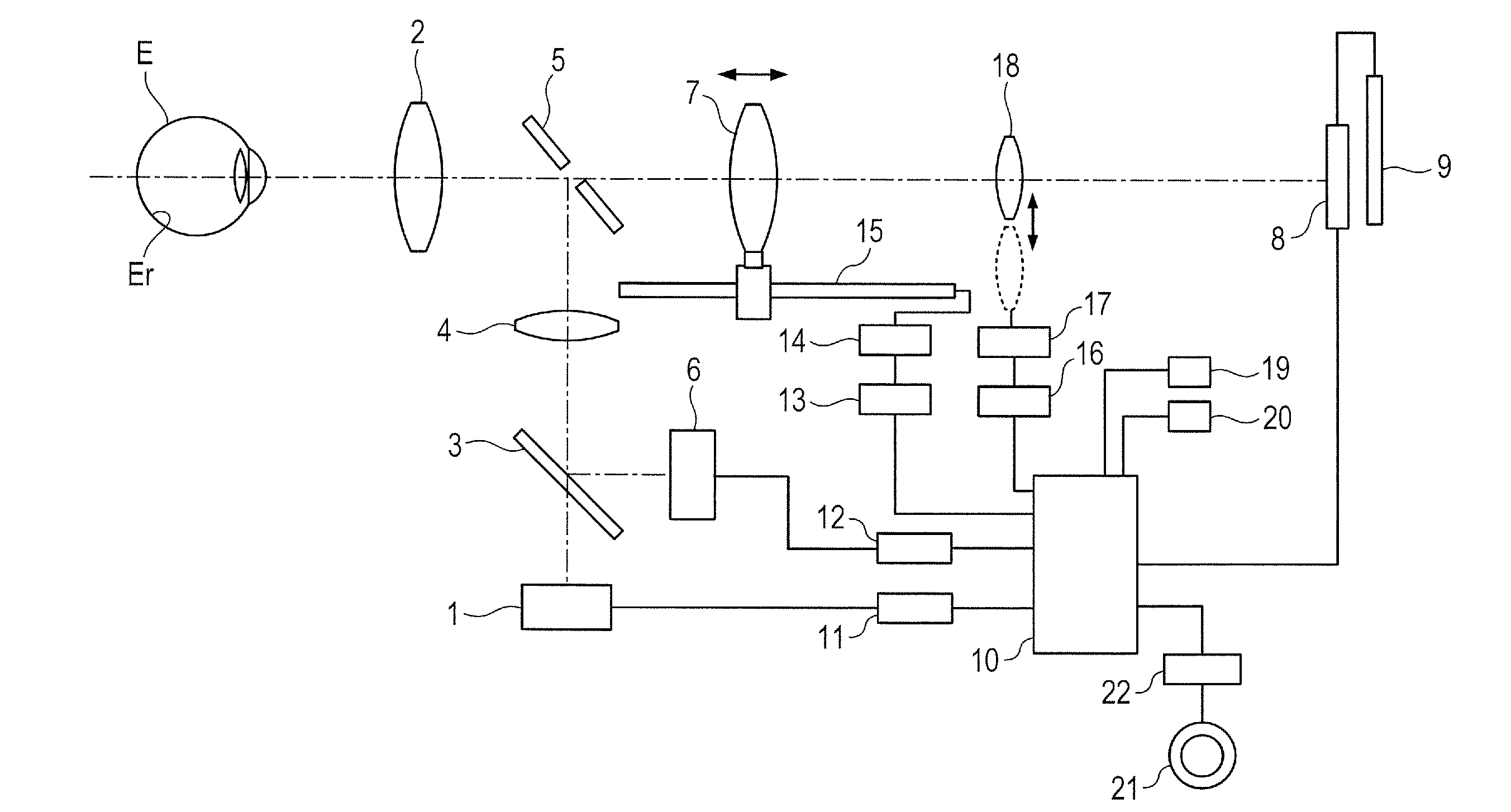
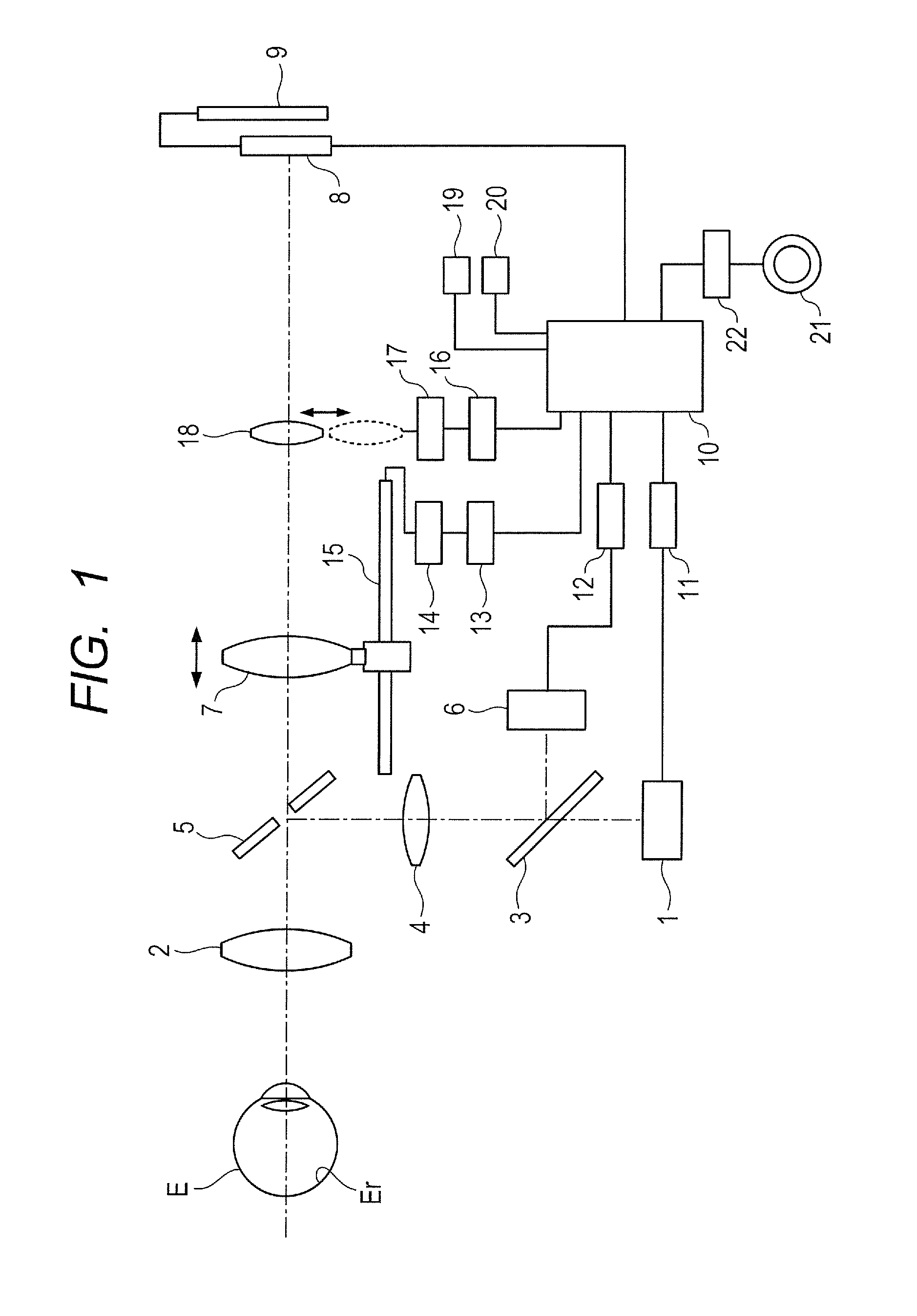
[0026]FIG. 1 is a view showing the arrangement of a fundus camera according to the first embodiment which is used as an ophthalmologic imaging apparatus. The fundus camera of this embodiment is a non-mydriatic fundus camera. The fundus camera located in front of an eye E to be inspected incorporates an observation illumination optical system ranging from an observation light source 1, which is formed from, for example f an infrared LED and emits infrared light, to an objective lens 2 located in correspondence with the eye E. In this observation illumination optical system, the observation light source 1, a dichroic mirror 3, a relay lens 4, and a perforated mirror 5 are sequentially arranged. An imaging light source 6 formed from a xenon tube is located as an imaging illumination optical system in the incident direction of the dichroic mirror 3.
[0027]A focus lens 7 which adjusts focus by moving in the optical axis direction is located as an imaging optical system behind the per...
second embodiment
[0059
[0060]The second embodiment of the present invention will be described next.
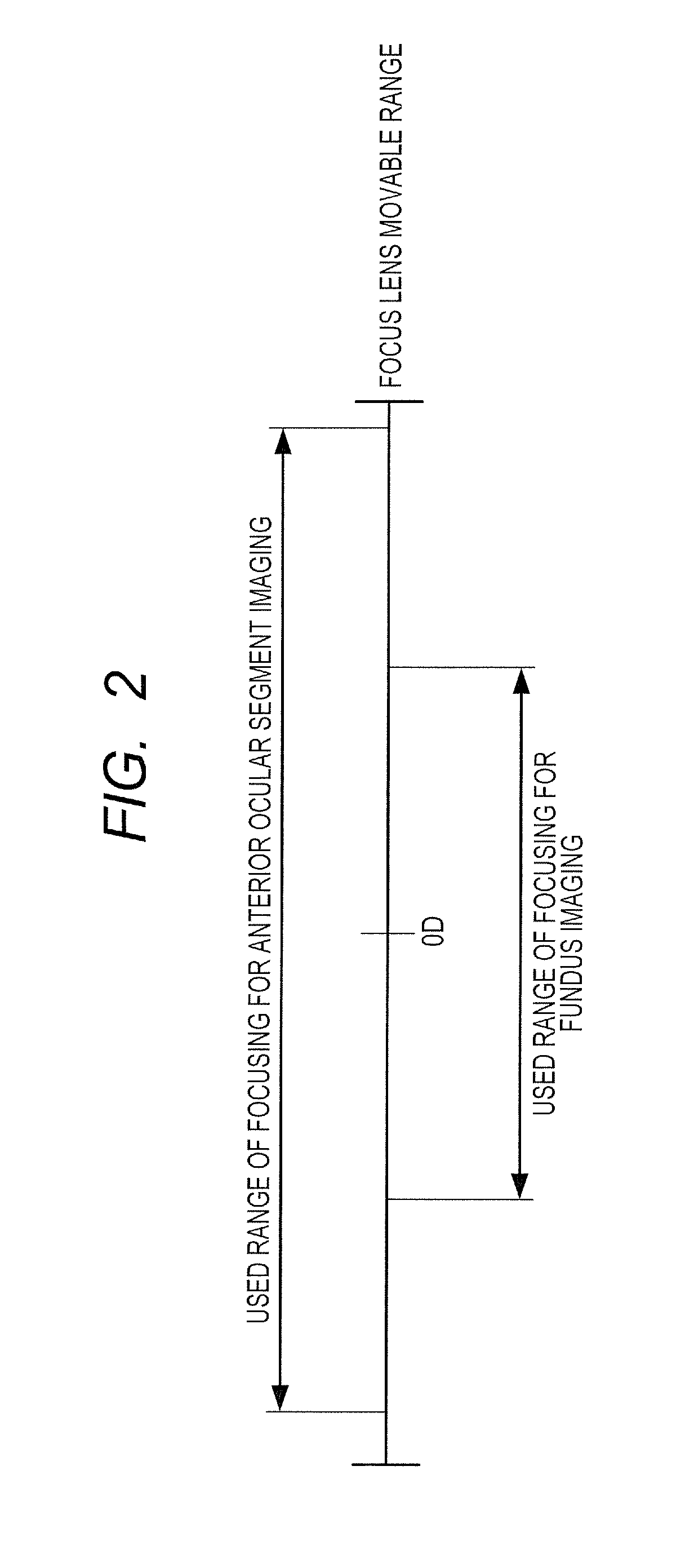
[0061]In anterior ocular segment imaging, operators differ in their regions of main interest, but a specific operator tends to perform imaging under predetermined conditions unique to him / her. In such a case, the operator does not always use the entire movable range of focusing for anterior ocular segment imaging, and hence allowing the operator to finely set a specific range of focusing will provide better usability.
[0062]The second embodiment has been made in consideration of this point and is configured to switch between at least two types of sensitivities for anterior ocular segment imaging operation, namely fine alignment and rough alignment.
[0063]The second embodiment will be described below with reference to FIG. 4. FIG. 4 is a view showing the arrangement of an ophthalmologic imaging apparatus according to the second embodiment. The same reference numerals as in FIG. 1 denote the same components...
third embodiment
[0075
[0076]The third embodiment of the present decent roc will be described next.
[0077]As described in the second embodiment, a specific operator tends to perform anterior ocular segment imaging under predetermined conditions unique to him / her. For this reason, enabling the operator to set a range of focusing for anterior ocular segment imaging by himself / herself can further improve the operabiiity. The third embodiment is configured to nave such an arrangement.
[0078]The third embodiment will be described below with reference to FIG. 7. FIG. 7 is a view showing the arrangement of an ophthalmologic imaging apparatus according to the third embodiment. The same reference numerals as in FIGS. 1 and 4 denote the same components in FIG. 7, and a description of them will be omitted.
[0079]This embodiment adds a focusing position set switch 24 for anterior ocular segment and a focusing position return switch 25 for anterior ocular segment to a control unit 10.
[0080]When the operator operates...
PUM
 Abstract
Description
Claims
Application Information
 - R&D
- Intellectual Property
- Life Sciences
- Materials
- Tech Scout
- Unparalleled Data Quality
- Higher Quality Content
- 60% Fewer Hallucinations
Browse by: Latest US Patents, China's latest patents, Technical Efficacy Thesaurus, Application Domain, Technology Topic, Popular Technical Reports.
© 2025 PatSnap. All rights reserved.Legal|Privacy policy|Modern Slavery Act Transparency Statement|Sitemap|About US| Contact US: help@patsnap.com


