
System for MRI-guided interventional mammary procedures

a technology of interventional procedures and magnetic resonance imaging, applied in the field of magnetic resonance imaging-guided interventional procedures, can solve the problems of inconvenient limited design of devices, and inability to provide compression along a specific orientation, and achieve accurate positioning and monitoring of interventional procedures, enhance the efficacy and efficiency of breast intervention, and enhance the effect of magnetic resonance imaging

Inactive Publication Date: 2005-06-07
RGT UNIV OF MINNESOTA
View PDF17 Cites 122 Cited by
  • Summary
  • Abstract
  • Description
  • Claims
  • Application Information

AI Technical Summary

Benefits of technology

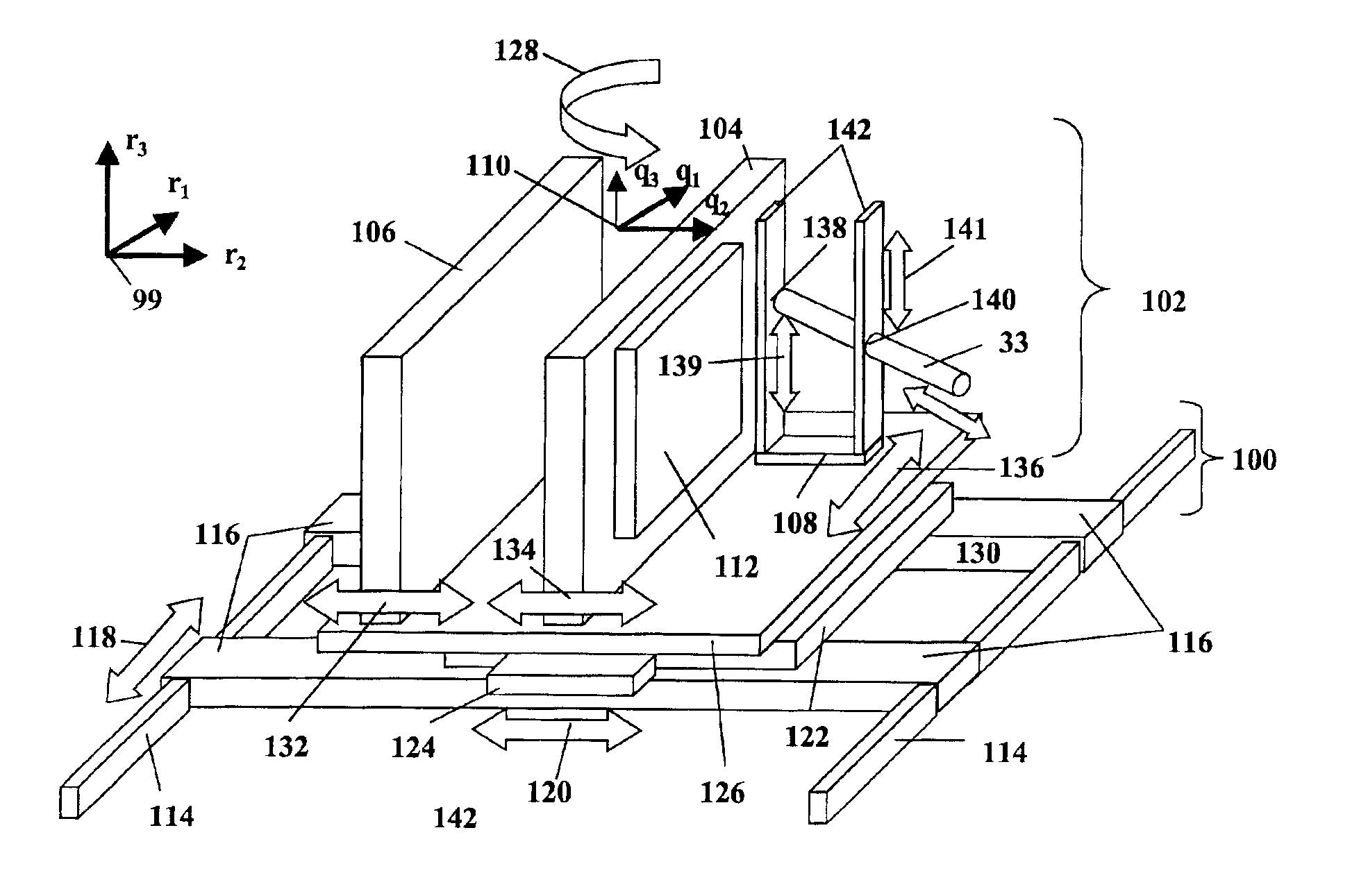
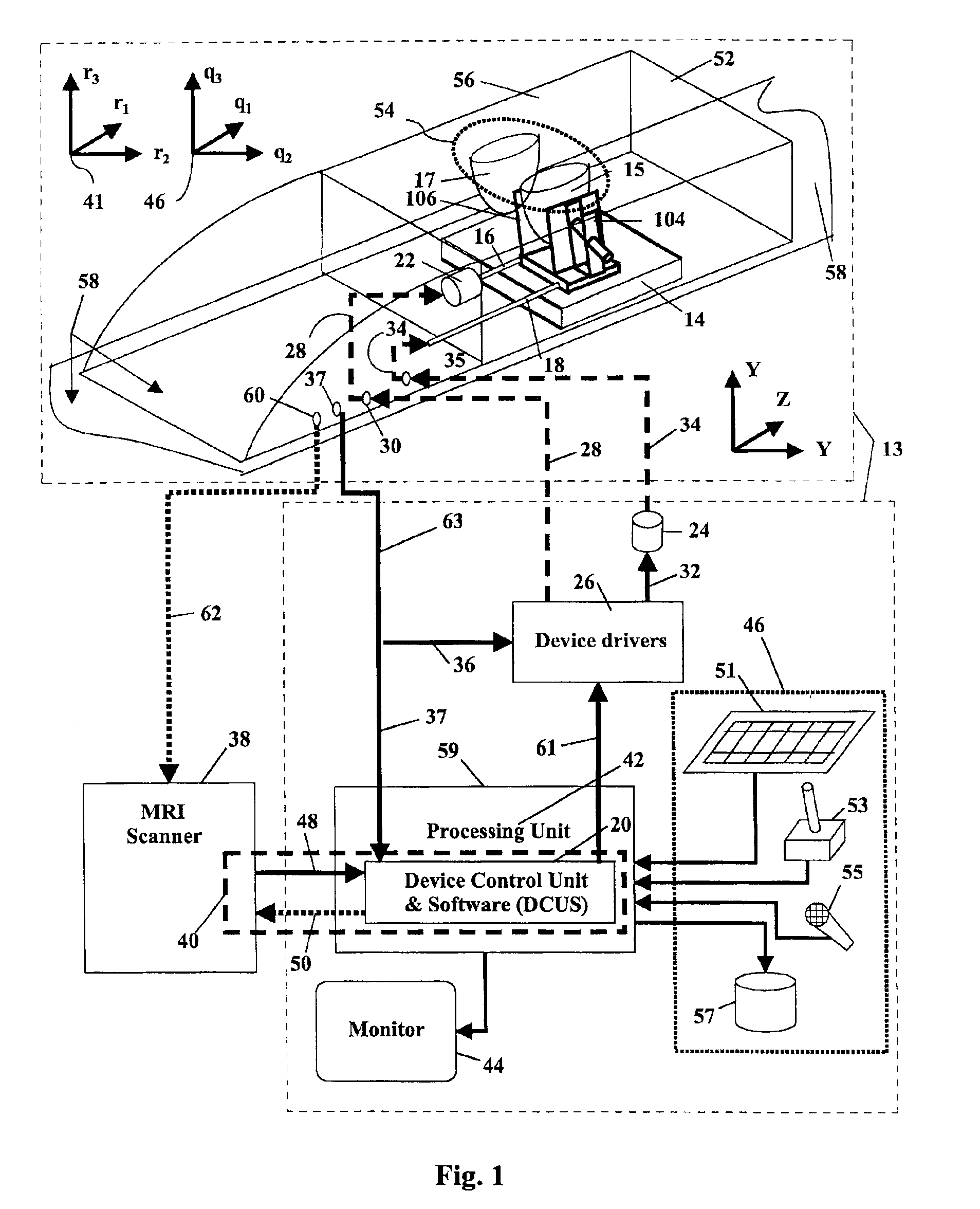
[0022]The combination of contrast enhanced magnetic resonance imaging (MRI) and MR-guided subcutaneous core biopsy can be used as a robust approach for the diagnosis and treatment of breast cancer. MRI provides the means to accurately position and monitor interventional procedures such as biopsy, removal of tissue or other transcanular procedures. MRI may also be used in this invention to position and monitor the progress of breast conserving therapies (BCT), such as laser photo-ablation, cryoablation and localized hyperthermia. The general practice of this invention is to provide a remotely controlled apparatus for MR-guided interventional procedures in the breast. The apparatus allows the practice of a method that provides flexibility in conditioning the breast, i.e. orientation and degree of compression, and in setting the trajectory of the intervention.
[0023]To that end, a robust conditioning / positioning device, fitted with the appropriate degrees of freedom, enhances the efficacy and efficiency of breast interventions by providing the flexibility in planning and executing an appropriate procedure strategy that better suits interventional procedures, either those in current use or yet to be developed. The novelty and potential commercial success of the device originates from its high maneuverability to set and perform the procedure strategy and its adaptability to accommodate an array of interventional probes. Remote control of this device can allow planning the operation and performing the relevant tasks in a short period, for example, within the contrast window provided by a single injection of a contrast agent, and this feature can be operator-independent.
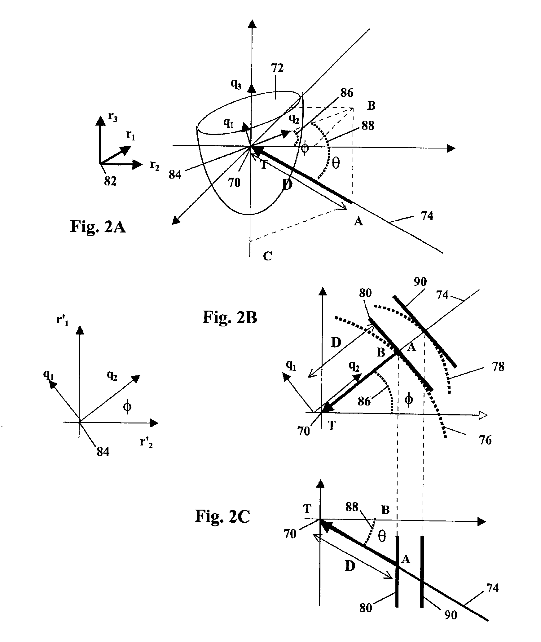
[0024]A remotely controlled device may be used that provides high accessibility to the target in the breast and a single step set-up of the procedures. The device may be equipped with a quadrature or linear RF coil with variable width. The remotely controlled device may allow for high flexibility in conditioning the breast and accessing a target inside a breast that better suits the interventional procedure, with the following features optimally used. For example the controller could benefit from: a) Operator-defined orientation and degree of compression and trajectory, i.e. orientation of probe insertion, for optimized access to any area in the breast and, in particular, for difficult-to-reach areas. b) A remote control mechanism of mechanical or hydraulic actuators and MR-visible markers for verifying and monitoring the execution of the planned operation with an MRI scanner. c) Suitable software for planning the intervention and operating the device. d) A generic instrumentation platform which can accommodate a variety of probes and accessories. e) Suitable degrees of freedom to accomplish the aforementioned task, and in particular its ability to position the intervention probes at any spatial position with given spatial limitations, as imposed by the wide variety of breast and chest anatomies and lesion positions that may be presented. f) The accuracy and repeatability to position the interventional probes at given spatial locations. g) Its compatibility for operation inside an MRI scanner. h) A suitable radio frequency (RF) coil such as a quadrature biplanar RF coil with adjustable spatial separation of the two planar elements to conform with the shape alterations of the compressed breast. The remotely controlled device may be moved by any convenient means. For example, there may be direct manual control, dials maneuvering the device through a mechanical interface, or computer driven controls that drive the device.
[0025]The general methodology of practice of the process in the present invention under MRI guidance comprises injecting a contrast agent into the patient at a position of injection that will enable the contrast agent to spread within tissues of the breast; allowing the contrast agent to spread into the tissues of the breast to reach at least a predetermined level of contrast; then condition the breast to restrict the flow of blood into and out of the breast to increase the persistence of the contrast agent within the breast. When that initial preparation of the area of the breast to be imaged has been performed, an operator may observe the breast under non-invasive procedures that use or require the use of contrast agents in the observational procedure. Then an interventional procedure may be used or performed under the observational technique while the contrast agent persists in the area of concern. This contrast persisting technique can increase the time that the procedure may be performed under adequate observational conditions, while minimizing the amount and number of times that contrast agent needs to be provided or injected.
[0026]This procedure increases the time under which the procedure may be performed, requires fewer and lower quantitative doses of contrast agent, requires fewer manipulations of the breast tissue while physically restricted, and generally makes non-interventional breast observation more tolerable to the patient.

Problems solved by technology

These findings result in a patient management dilemma when lesions are detected with MRI and those lesions have not been seen with other gold standard modalities.
Despite their success, there are some limitations in these designs, particularly when they are compared with the non-MR stereotaxic devices.
First, most of the devices provide compression along a specific orientation, usually medial-lateral, which may not be the optimal one, as for example for transversing the shortest path in the tissue or to reach areas such as the axilla.
Second, in most of the devices the probe is directed by means of a mesh, and thus it can be only inserted perpendicular to the compression plates or with a slight “free-hand” angulation.
This may not be the optimal approach, for example, when attempting to access tissue close to the chest wall, at the axilla or to avoid obstructions such as implants.
The two single plate systems have the same limitations and in addition there is the potential for accidental rib puncture or highly invasive operations behind the nipples.
This practice increases the length of the operation, and may not be always feasible due to the limited number of allowed injections of contrast material and the short duration of the contrast window.
These RF coils, except for those shown in Greenstein-Orel, et al. and E. K. Insko, et al., are of standard dimensions and are not appropriate for use with the proposed apparatus since they will have a variable filling-factor for different degrees of breast compression.
While endoscopic, arthroscopic, and endovascular therapies have already produced significant advances in health care, these techniques ultimately suffer from the same limitation.
This limitation is that the accuracy of the procedure is “surface limited” by what the surgeon can either see through the device itself or otherwise visualize (as by optical fibers) during the course of the procedure.
That is, the visually observable field of operation is quite small and limited to those surfaces (especially external surfaces of biological masses such as organs and other tissue) observable by visible radiation, due to the optical limitations of the viewing mechanism.
At the same time, the limited distribution of drug injected from a single catheter tip presents other problems.
Delivery of a drug from a single point source may also limit the distribution of the drug by decreasing the effective radius of penetration of the drug agent into the surrounding tissue receptor population.
However, there is significant pain and discomfort involved in the present procedures that causes many patients to resist regular and early examination.

Method used


Image

  • System for MRI-guided interventional mammary procedures
  • System for MRI-guided interventional mammary procedures
  • System for MRI-guided interventional mammary procedures

Examples


Embodiment Construction

[0045]The general methodology of practice of the process in the present invention comprises injecting a contrast agent into the patient at a position of injection that will enable the contrast agent to spread within tissues of the breast; allowing the contrast agent to spread into the tissues of the breast to reach at least a predetermined level of contrast; then condition the breast to restrict the flow of blood into and out of the breast to increase the persistence of the contrast agent within the breast. When that initial preparation of the area of the breast to be imaged has been performed, an operator may observe the breast under non-invasive procedures that use or require the use of contrast agents in the observational procedure. Then an interventional procedure may be used or performed under the observational technique while the contrast agent persists in the area of concern. This contrast persisting technique can increase the time that the procedure may be performed under ad...


PUM


Abstract

The combination of contrast enhanced magnetic resonance imaging (MRI) and MR-guided subcutaneous core biopsy can be used as a robust approach for the diagnosis and treatment of breast cancer. MRI provides the means to accurately position and monitor interventional procedures such as biopsy, removal of tissue or other transcanular procedures. MRI may also be used in this invention to position and monitor the progress of breast conserving therapies (BCT), such as laser photo-ablation, cryoablation and localized hyperthermia. The general practice of this invention is to provide a remotely controlled apparatus for MR-guided interventional procedures in the breast. The apparatus allows the practice of a method that provides flexibility in conditioning the breast, i.e. orientation and degree of compression, and in setting the trajectory of the intervention. To that end, a robust conditioning / positioning device, fitted with the appropriate degrees of freedom, enhances the efficacy and efficiency of breast interventions by providing the flexibility in planning and executing an appropriate procedure strategy that better suits interventional procedures, either those in current use or yet to be developed. The novelty and potential commercial success of the device originates from its high maneuverability to set and perform the procedure strategy and its adaptability to accommodate an array of interventional probes. Remote control of this device can allow planning the operation and performing the relevant tasks in a short period, for example, within the contrast window provided by a single injection of a contrast agent, and this feature can be operator-independent.

Description

CLAIM OF PRIORITY[0001]This application is a divisional of U.S. patent application Ser. No. 09 / 407,346, filed Sep. 29, 1999, now U.S. Pat. No. 6,675,037, herein incorporated by reference.GOVERNMENT FUNDING[0002]The invention described herein was made with Government support by the National Institute of Health (NIH) under grant number R41CA81817 (National Cancer Institute). The United States Government has certain rights in the invention.BACKGROUND OF THE INVENTION[0003]1. Field of the Invention[0004]The present invention relates to Magnetic Resonance Imaging-Guided (MRI-Guided) medical procedures, particularly MRI-Guided interventional procedures and therapies that are performed on the breasts or mammaries of patients.[0005]2. Background of the Art[0006]The diagnosis and treatment of breast cancer is a major health care issue which affects the lives of more than 180,000 women annually, only considering the United States. While, early detection and treatment of breast cancer is a maj...

Claims


Application Information

Patent Timeline
IPC IPC(8): A61B19/00A61B5/055A61B10/00A61B10/02
CPCA61B5/0555A61B10/0233A61B19/20A61B10/0041A61B2019/5458A61B19/22A61B2019/205A61B2019/5236A61B19/201A61B90/10A61B90/17A61B34/70A61B90/11A61B2090/374A61B2090/3958A61B5/055A61B5/708
Inventor TSEKOS, NIKOLAOS V.
Owner RGT UNIV OF MINNESOTA
Learn More