Apparatus and method for an inclined single plane imaging microscope box (ispim box)

a single-plane imaging and microscope box technology, applied in the field of single-plane illumination microscopy, can solve the problem of lack of opm limitation, which requires the construction of a new microscope body,

Active Publication Date: 2014-10-30
RGT UNIV OF CALIFORNIA
View PDF16 Cites 12 Cited by
  • Summary
  • Abstract
  • Description
  • Claims
  • Application Information

AI Technical Summary

Benefits of technology

[0016]The illustrated embodiments of the invention are directed to a variant of SPIM that has all the advantages of the SPIM technology including that it can operate on samples prepared with standard protocols including dish cell cultures. In illustrated embodiments of the invention we disclose a SPIM system in which the light sheet is produced at an angle. Our setup can be installed as an add-on into any standard commercial microscope. It uses only one objective lens in proximity of the sample and a second objective along the emission path. These embodiments allow us to use standard modalities for sample preparation and insertion while the SPIM effect is done elsewhere in the microscope, far from the sample. Since the light goes through the sample at an angle, we called these embodiments the iSPIM (inclined SPIM).
[0017]The main use of the iSPM Box of the illustrated embodiments is microscopy imaging, in particular for three dimensional volumetric acquisition. It is also possible to perform fluorescence correlation spectroscopy (FCS) on the images acquired. The inclined single plane illumination microscopy (iSPIM) box is a single-sided add-on module that allows user to perform iSPIM on virtually any microscope body. The system is intended for imaging biological samples with a fast volumetric acquisition to produce three dimensional images. It finds application in any bioimaging related field that makes use of fluorescence for imaging. iSPIM is based on the single plane illumination microscopy principle. It uses a cylindrical lens to create a thin laser sheet which optically sections the sample with high efficiency of light illumination and detection. As a consequence of this high efficiency the sample undergoes a consistently reduced photo-bleach and energy load. The acquisition speed of the system is the highest among microscopy systems.
[0018]In the illustrated embodiments of the invention we incorporate all the optics necessary for the OPM in a single box that can be used as an add-on to existing commercial microscopes. We named this design the inclined single plane illumination microscopy box (iSPIM box). It maintains the same advantage of OPM over original SPIM which are: conventional sample preparation techniques. e.g. glass microscope slides and cell culture dishes may be used; minimal photobleaching, phototoxicity and energy load of the sample; no moving parts required to obtain a two dimensional image-good for imaging dynamics; no calculation required to get sectioned image; good three dimensional imaging when combined with a motorized xy-stage to position the sample

Problems solved by technology

The limitation of OPM which requires the construction of a new microscope body is lacking in the iSPIM box construction.

Method used


Image

  • Apparatus and method for an inclined single plane imaging microscope box (ispim box)
  • Apparatus and method for an inclined single plane imaging microscope box (ispim box)
  • Apparatus and method for an inclined single plane imaging microscope box (ispim box)

Examples


Embodiment Construction

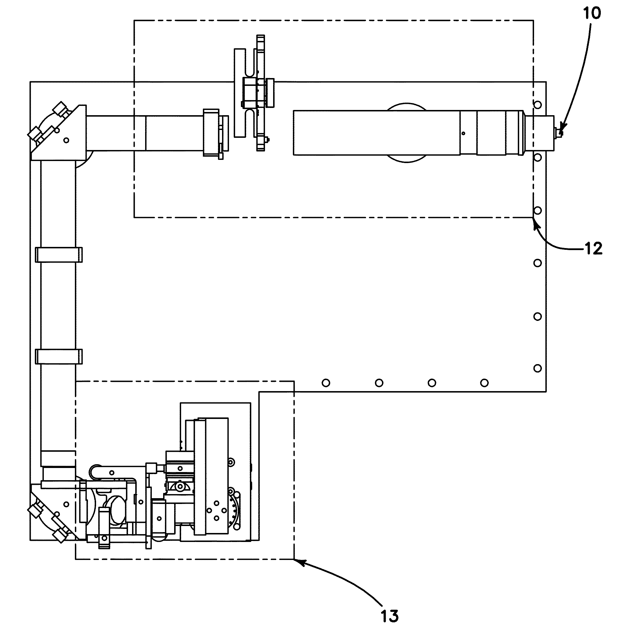
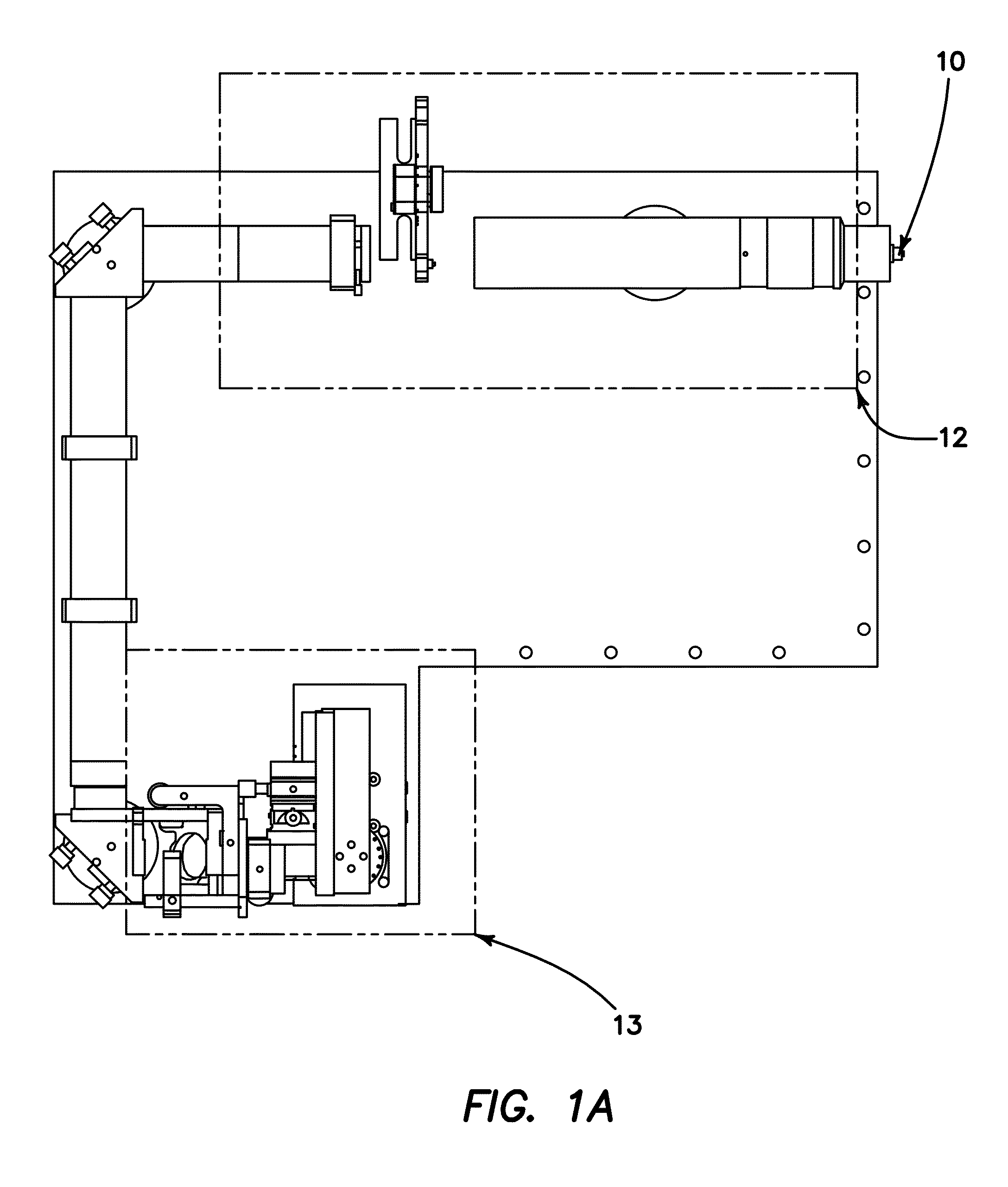
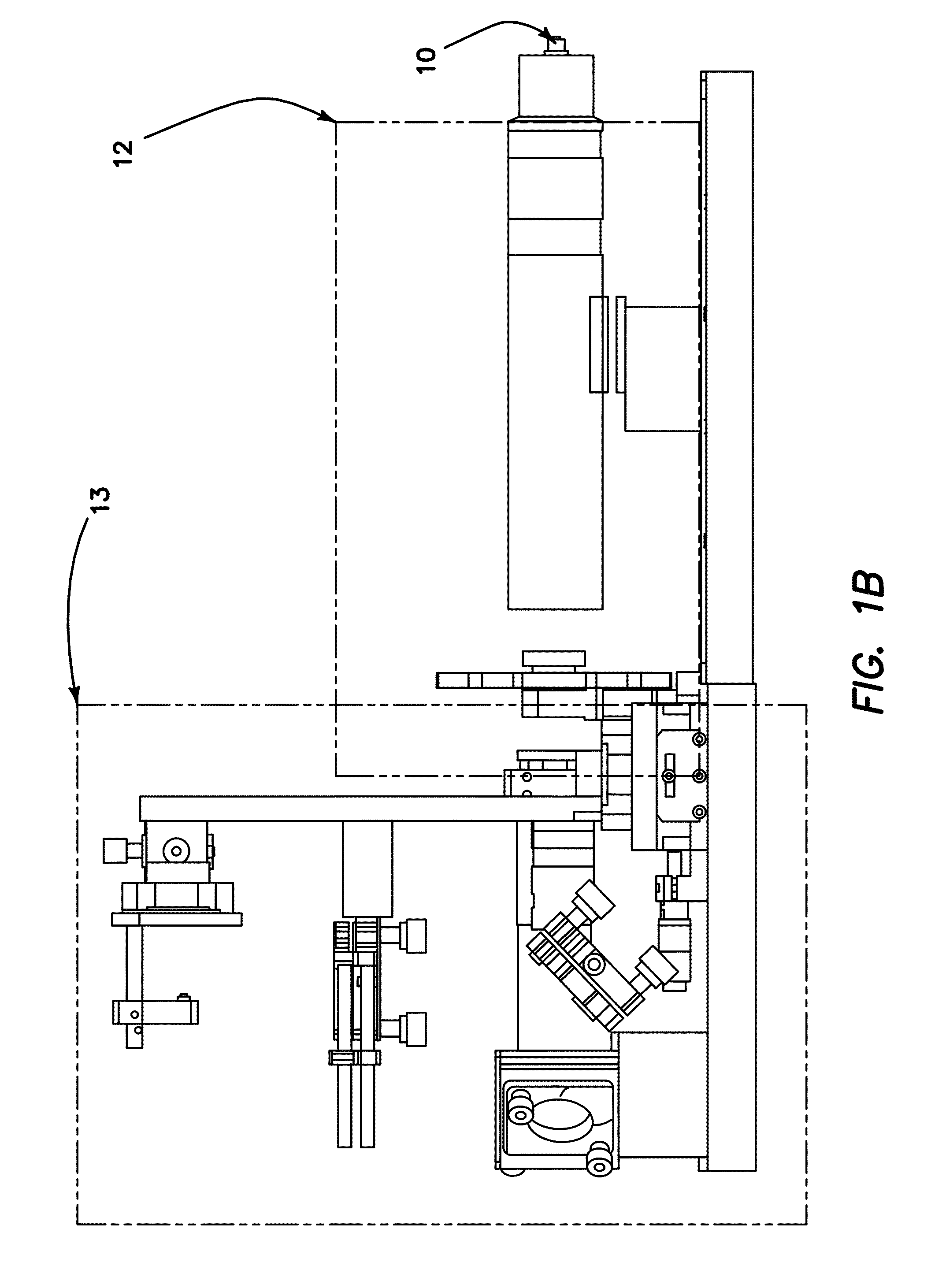
[0046]The disclosed system for inclined selective plane illumination microscope (iSPIM) with shares the advantages of OPM and that can be used as an add-on to commonly used microscopes, such as a conventional IX-71 Olympus microscope, simplifying the construction of the OPM and increasing performance of a conventional microscope. As shown below in the diagrammatic depictions of FIGS. 1a-1d the primary components include: a laser input connector or laser 10, a beam expanding component 12, an universal dichroic component 13 to separate fluorescence from excitation light, an universal optical adaptor 14, a re-imaging component 15, and a camera output connector 16.

[0047]FIG. 1d is a typical setup of an iSPIM. Laser beam from laser 10 is inserted into a 10× beam expander 12 and reflected by a mirror 14 both mounted on a vertically moving component 17. Cylindrical lens 18 and achromatic doublet 20 are mounted on the “injection arm”22. The dichroic mirror 13 reflects the beam vertically in...


PUM


Abstract

An apparatus for inclined single plane Illumination microscopy of a sample includes a laser for launching excitation light beams at a plurality of wavelengths, a laser beam expander, an injection arm optically coupled to the laser beam expander, a conventional back-to-back microscope system, a universal dichroic mirror optically coupled to the injection arm to direct the excitation light beams into the conventional back-to-back microscope onto a sample plane in an imaging plane, and to receive fluorescence light from the sample, a universal optical adaptor optically coupled to the universal dichroic mirror, a re-imaging component optically coupled to the universal optical adaptor; and a camera output connector optically coupled to the re-imaging component, where the laser beam expander, injection arm, universal optical adapter, re-imaging component, and camera are combined in a modular unit which is arranged and configured to be coupled to the conventional back-to-back microscope.

Description

GOVERNMENT RIGHTS[0001]This invention was made with Government support under grant nos. GM076516 and RR003155 awarded by the Institutes of Health. The Government has certain rights in this invention.BACKGROUND[0002]1. Field of the Technology[0003]The disclosure relates to the field of single plane Illumination microscopy (SPIM).[0004]2. Description of the Prior Art[0005]The need for fast three dimensional microscopy has increased over the years in bio-imaging facilities around the world. In order to respond to this need, use of the confocal microscope principle has been pushed to faster speed such as the spinning disk technology (Nipkow disk optimizations) and fast laser scanning microscopes.[0006]Recently, another technique has been proposed, selective plane illumination microscopy (SPIM), a method based on illumination of the sample from the side with a sheet of light. The original idea was described in 1903 (Siedentopf and Zsigmondy, 1903) and pioneered in 1993 (Voie et al., 1993...

Claims


Application Information

Patent Timeline
Patent Type & Authority Applications(United States)
IPC IPC(8): H04N13/02
CPCH04N13/02G02B21/16G02B2207/113
Inventor CUTRALE, FRANCESCOGRATTON, ENRICO
Owner RGT UNIV OF CALIFORNIA
Why Eureka
Try Eureka