Motion-tracking x-ray ct image processing method and motion-tracking x-ray ct image processing device

Inactive Publication Date: 2014-11-13
KYOTO UNIV
View PDF6 Cites 9 Cited by
  • Summary
  • Abstract
  • Description
  • Claims
  • Application Information

AI Technical Summary

Benefits of technology

The patent describes a way to process X-ray CT images to reduce motion artifacts. This is achieved by estimating the motion and the CT image simultaneously, which helps to smoothly deform the measuring target and eliminate the need for synchronization with an electrocardiogram. This method can also be used to estimate the motion of internal organs that cannot be synchronized with an electrocardiogram. Additionally, this approach allows for real-time estimation, which can reduce the exposure dose to the patient.

Problems solved by technology
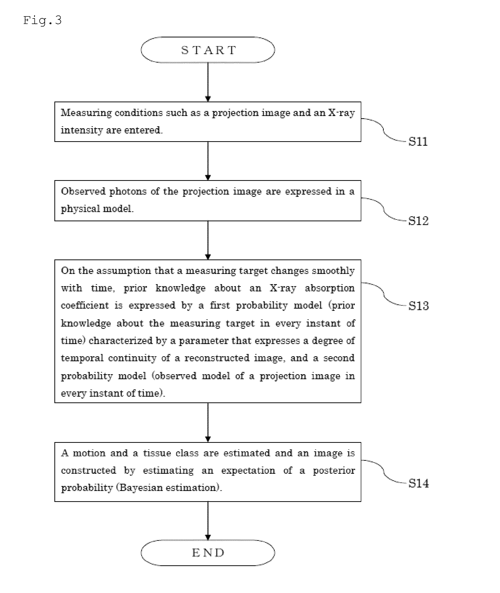
However, objects targeted for X-ray CT for medical purposes include hearts, blood vessels, lungs and infants that find difficulty in stopping their motions completely.
Applying an X-ray CT algorithm assuming a standstill to such active objects like in a conventional way is known to generate false images called motion artifacts.

Method used


Image

  • Motion-tracking x-ray ct image processing method and motion-tracking x-ray ct image processing device
  • Motion-tracking x-ray ct image processing method and motion-tracking x-ray ct image processing device
  • Motion-tracking x-ray ct image processing method and motion-tracking x-ray ct image processing device

Examples


embodiment 1

[0129]In Example 1, an object to be deformed was formed artificially and a screen was reconstructed based on a projection image of the object by calculator simulation using the motion tracking X-ray CT image processing method of the present invention. A result of the simulation is given below.

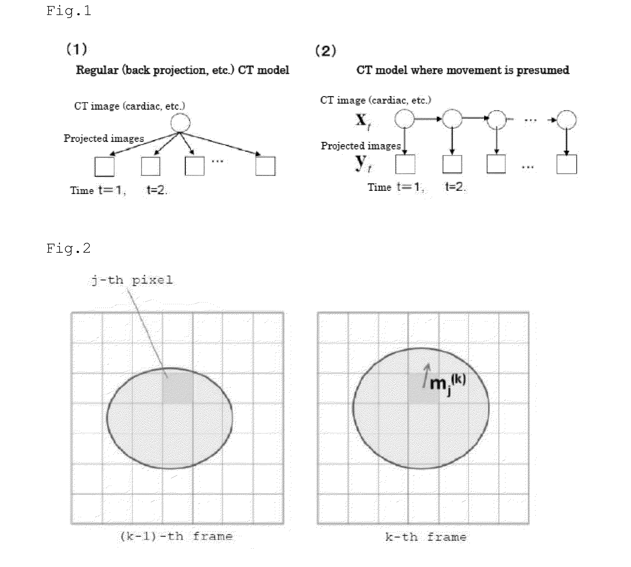
[0130]Images in FIG. 8 show an artificially formed image to be deformed. As shown in FIG. 8, a central area of each round screen shows the object (64×64 pixels). The object expanded from (a) to (e) with time. The deformation of the object is such that the object expanded at a rate differing between the vertical and horizontal directions.

[0131]FIG. 9 shows results of reconstruction of X-ray CT images. FIG. 9 is given to compare true images, results of image reconstruction obtained by a filtered back projection (FBP) method employed most frequently as a conventional method not assuming a motion, and results obtained by the image reconstruction of the present invention (suggested method).

[0132]Dif...

embodiment 2

[0138]The motion tracking X-ray CT image processing method of the present invention uses a probability model that makes statistical estimation as described above, so that it can incorporate other prior knowledge easily and can increase the quality of image reconstruction. As an example, making estimation in consideration of X-ray absorption coefficients representative of each tissue can enhance estimation accuracy. In this case, incorporating a hidden state variable to control the distribution of each tissue class can enhance estimation accuracy and can achieve robust estimation. The following describes experiment conducted to check the effect thereof.

[0139]FIG. 12 shows the X-ray absorption coefficient of an assumed tissue class and the value of the X-ray absorption coefficient of an object actually used in experiment. Here, it is assumed that there are four tissue classes including air, fat, muscle and bone, and that there is X-ray absorption coefficients representative of each ti...

embodiment 3

[0144]In Example 3, an X-ray CT algorithm is described different from that of Examples 1 and 2 described above.

[0145](1) Probability Model

[0146](1-a) Likelihood

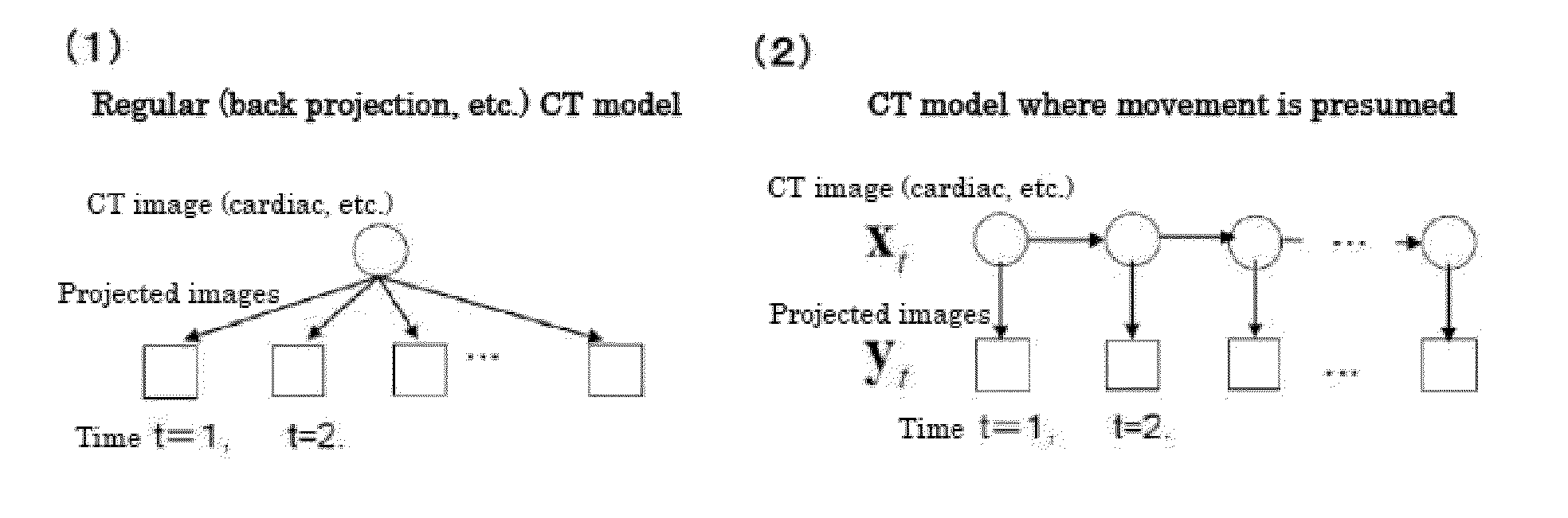
[0147]ACT image of an object targeted for reconstruction is expressed by an one-dimensional vector X=[x1, . . . , xJ] (J is the number of pixels of a reconstructed image). Projection mentioned herein is to be made on an object to be deformed or to move with time. Accordingly, time is divided into K frames S{k}, kε{1, . . . , K} and a CT image in a k-th frame is expressed as X{k}=[x1{k}, . . . , xJ{k}]. CT images X{k} (kε{1, . . . , K}) in all the frames are expressed collectively as X={X{1}, . . . , X{k}}.

[0148]An image obtained by projection on the object at a time t is expressed as Y{t}=[y1{t}, . . . , y1{t}] (I is the number of detectors) and projections in all instants of time are expressed collectively as D={Y{1}, . . . , Y{T}}. It is assumed that projection of a CT image X(k) is obtained if the time t satisfies tεS(k). ...


PUM


Abstract

An x-ray CT image processing method which carries out a statistical estimate in prior knowledge relating to the movement and the x-ray absorption coefficient of a measurement subject comprises: a step of hypothesizing that the measurement subject changes smoothly over time, and defining a first probability model (prior knowledge of the measurement subject over all times) relating to movement and a second probability model (projected image observation model over all times) relating to observation; and a step of carrying out a statistical estimate dependent on both the first probability model and the second probability model. The first probability model relating to the movement of the measurement subject and the second model relating to observation are defined as probability models with which statistical estimates are carried out at the outset, and motion and CT images are simultaneously estimated by carrying out induction based on the probability models.

Description

TECHNICAL FIELD[0001]The present invention relates to an X-ray CT algorithm capable of tracking a motion of a measuring target, a program having the algorithm, and an X-ray CT device on which the program is installed.BACKGROUND ART[0002]An X-ray CT is a powerful technique capable of acquiring an internal structure in a noninvasive and nondestructive manner and is being used for a wide range of medical purposes and industrial purposes such as defect inspection. The X-ray CT has been used for reconstructing an internal structure (CT image) based on the assumption that an object targeted for acquisition of its internal structure is at a standstill. However, objects targeted for X-ray CT for medical purposes include hearts, blood vessels, lungs and infants that find difficulty in stopping their motions completely. Applying an X-ray CT algorithm assuming a standstill to such active objects like in a conventional way is known to generate false images called motion artifacts.[0003]Cardiac ...

Claims


Application Information

Patent Timeline
IPC IPC(8): G06T11/00G06T7/20A61B6/03G06T7/00
CPCG06T11/008G06T7/0012G06T7/20A61B6/032G06T2207/10081G06T2207/30048A61B6/503A61B6/5258G06T11/005G06T2211/412G06T2207/20076G06T7/277
Inventor ISHII, SHINMAEDA, SHINICHITANAKA, TAKUMI
Owner KYOTO UNIV
Why Eureka
Try Eureka