
System and method for imaging a patient user's body part

Inactive Publication Date: 2014-11-27
NEUROVISION IMAGING INC
View PDF9 Cites 35 Cited by
  • Summary
  • Abstract
  • Description
  • Claims
  • Application Information

AI Technical Summary

Benefits of technology

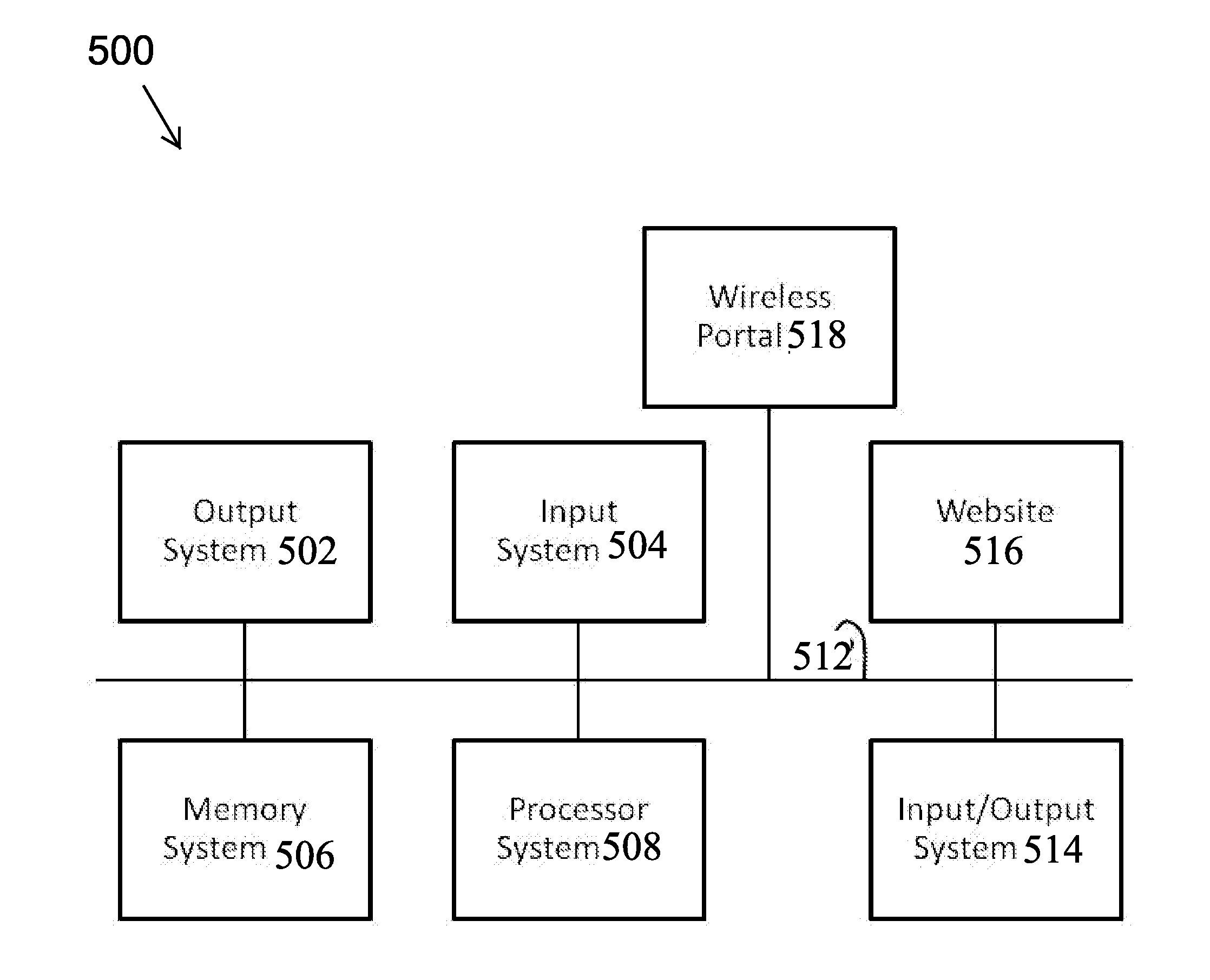
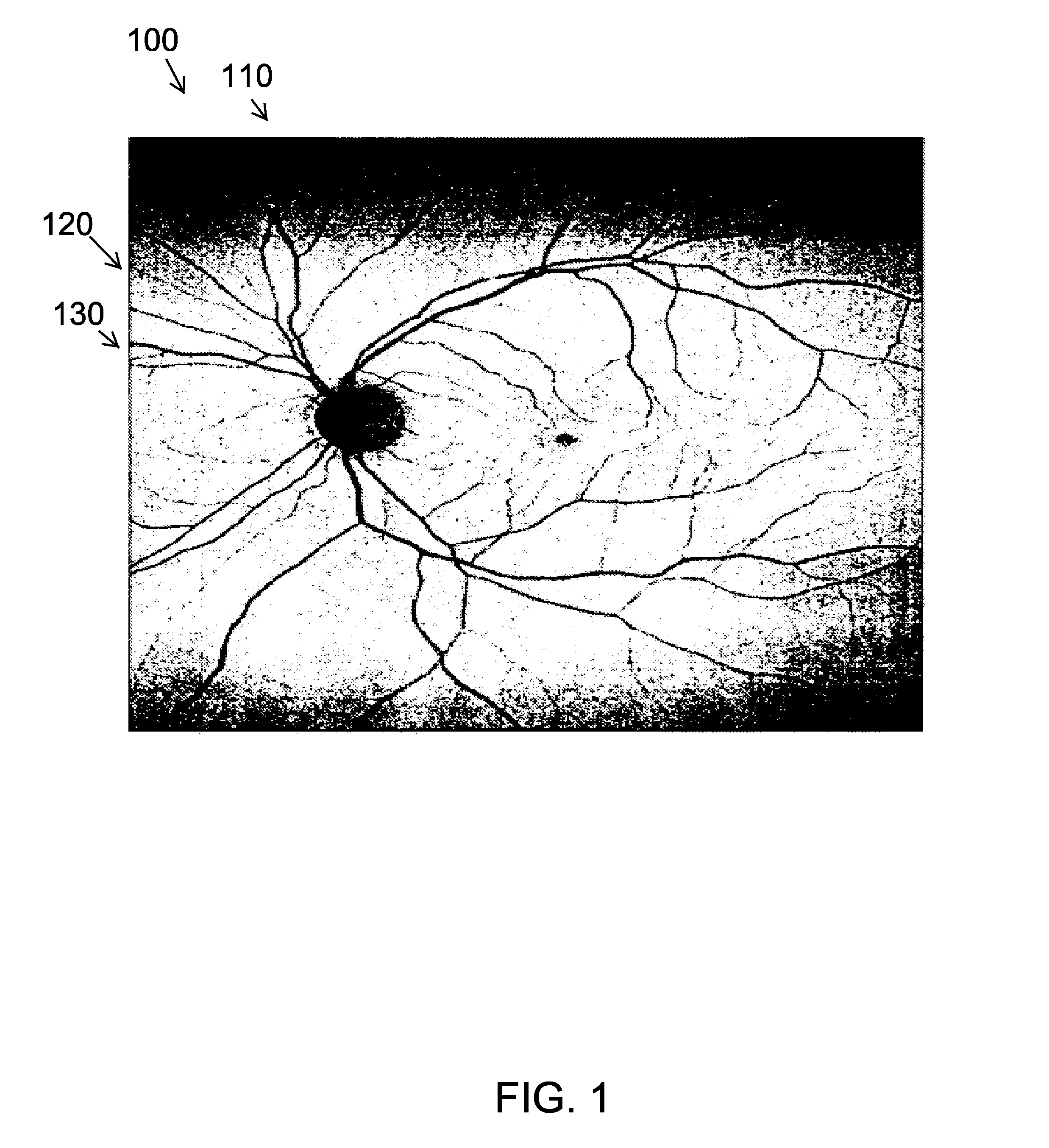
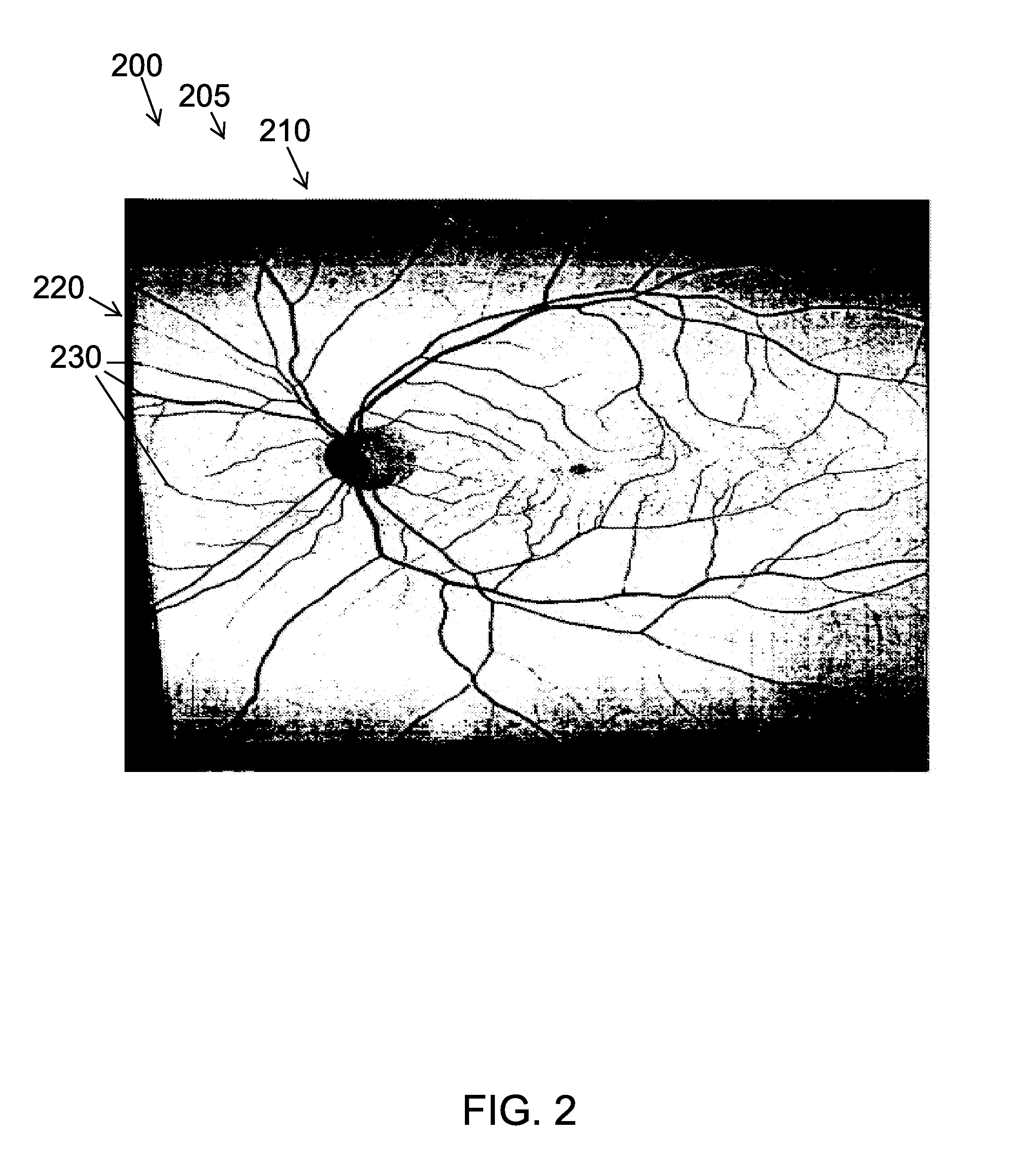
This patent describes a system and method for imaging a patient's body part. Unlike traditional imaging devices, this system can capture multiple in-focus regions of the eye, retina, ear, nose, throat, skin, and other body parts using a combination of packaging, optics, and image analysis. By recording movie streams and parsing good quality images from each frame, the overall resolution and image quality is greatly improved. This system is also more portable, affordable, and user-friendly than other imaging devices.

Problems solved by technology

The system and method for imaging a patient user's body part differs from other systems and methods in that traditional imaging devices do not afford for the visualization of multiple in-focus regions of the eye, retina, ear, nose, throat, skin or other interior or exterior body part, are not driven by SMARTPHONE® or tablet computer and are relatively large, expensive and cumbersome.

Method used


Image

  • System and method for imaging a patient user's body part
  • System and method for imaging a patient user's body part
  • System and method for imaging a patient user's body part

Examples


Embodiment Construction

[0041]Various aspects of the illustrative embodiments will be described using terms commonly employed by those skilled in the art to convey the substance of their work to others skilled in the art. However, it will be apparent to those skilled in the art that the present invention may be practiced with only some of the described aspects. For purposes of explanation, specific numbers, materials and configurations are set forth in order to provide a thorough understanding of the illustrative embodiments. However, it will be apparent to one skilled in the art that the present invention may be practiced without the specific details. In other instances, well-known features are omitted or simplified in order not to obscure the illustrative embodiments.

[0042]Various operations will be described as multiple discrete operations, in turn, in a manner that is most helpful in understanding the present invention. However, the order of description should not be construed as to imply that these op...


PUM


Abstract

A system and method for imaging a patient user's body part is disclosed. The system may include a patient user body part imaging non-transitory storage media to image a patient user body part. The method may include selecting an optical imaging device to image the patient user's body part, acquiring one or more data sets with the optical imaging device, the acquiring is performed with focus at multiple axial positions and exposure control or deliberate focus at specified image locations and exposure control, registering the acquired data sets, performing image processing on the acquired data sets and recombining good data from the image processed data sets into a single image of the patient user's body part.

Description

[0001]This application claims priority to U.S. Provisional Application 61 / 825,830 filed on May 21, 2013, the entire disclosure of which is incorporated by reference.BACKGROUND OF THE INVENTION[0002]1. Field of the Invention[0003]The present invention is a system and method for imaging. More specifically, the present invention is a system and method for imaging a patient user's body part.[0004]2. Description of the Related Art[0005]Imaging of a patient user's body part is typically done with one or more slit lamps, ophthalmoscopes, fundus cameras, scanning laser ophthalmoscopes or SLO's and wide field devices for imaging the eye, an otoscope for imaging the ear, nose or throat, a dermascope for imaging the skin, or an endoscope for imaging an interior organ or body cavity. These devices are often relatively expensive and require one or more personal computers, cameras, sensors and monitors. They are typically relatively large, require an instrument table, are not portable, are not ba...

Claims


Application Information

Patent Timeline
IPC IPC(8): A61B5/00
CPCA61B5/0059A61B5/0022A61B1/00A61B3/102G16H40/67
Inventor VERDOONER, STEVEN ROGER
Owner NEUROVISION IMAGING INC
Learn More