Apparatus for optical analysis of an associated tissue

an associated tissue and optical analysis technology, applied in the field of apparatus for optical analysis of associated tissue, can solve problems such as insufficientness

Inactive Publication Date: 2014-12-25
KONINKLJIJKE PHILIPS NV
View PDF7 Cites 4 Cited by
  • Summary
  • Abstract
  • Description
  • Claims
  • Application Information

AI Technical Summary

Benefits of technology

[0006]In particular, it may be seen as an object of the present invention to provide an apparatus, a method and a computer program which could be beneficial for discr

Problems solved by technology

Although differences in blood content will likely provide possibilities to discriminate certain struct

Method used


Image

  • Apparatus for optical analysis of an associated tissue
  • Apparatus for optical analysis of an associated tissue
  • Apparatus for optical analysis of an associated tissue

Examples


Embodiment Construction

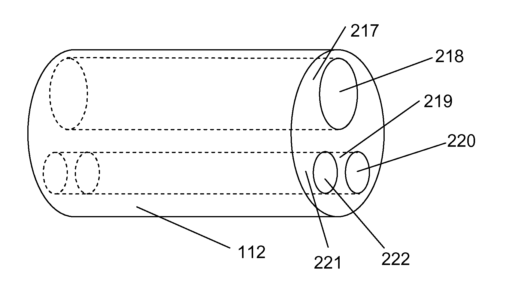
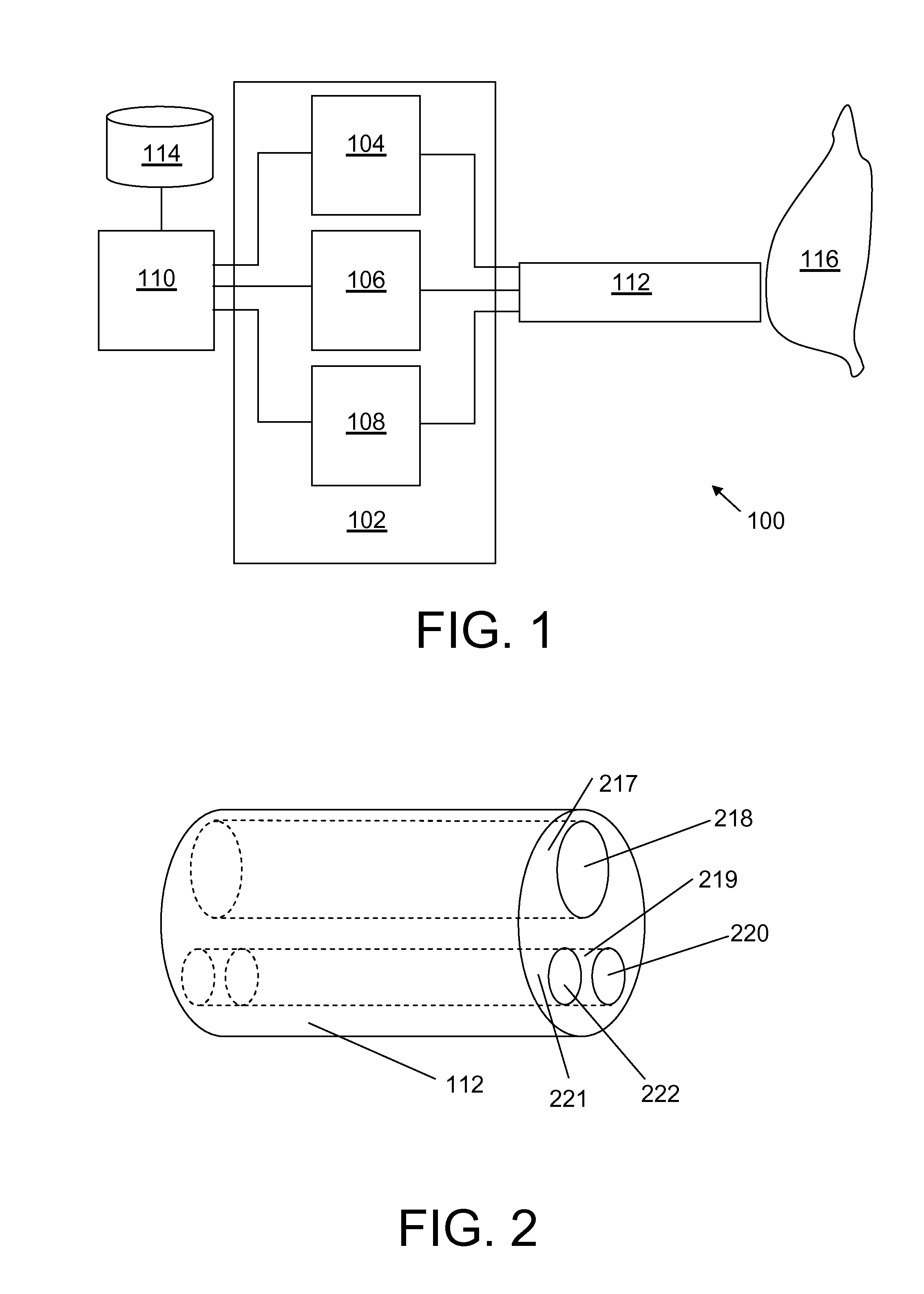
[0080]FIG. 1 shows a diagrammatic depiction of an apparatus according to an embodiment of the invention comprising a spectrometer 102 comprising a light source 104, a first optical detector 106, an optional second optical detector 108 and an interventional device 112, where the interventional device 112 has one or more guides, such as optical elements, such as optical waveguides, capable of guiding light from the light source 104 to a distal end of the interventional device so as to emit the light at the distal end of the interventional device, and furthermore capable of guiding light back from the distal end of the interventional device to the first optical detector 106 and / or second optical detector 108. The light guides enable light to enter an associated tissue 116, such as a prostate tissue, and the light guides further enable light exiting the associated tissue to be collected and led to the optical detector. The apparatus thus enables procurement of measured data representati...


PUM


Abstract

The present invention relates to an apparatus 100 and, a method and a computer program for determining a first parameter indicative of a concentration of lycopene. In particular, the invention relates to an apparatus 100 comprising a spectrometer 02, which spectrometer comprises a light source 104 and a detector 106, 108 arranged to measure an optical spectrum via an interventional device 112. This enables determination of a first parameter being indicative of a lycopene concentration. Lycopene concentration may serve as a discriminative feature for different tissue types, such as the prostate organ and associated structures. The apparatus may in a specific embodiment be arranged to determine a second parameter indicative of a tissue type based on a concentration of lycopene. According to a specific embodiment, the apparatus relies on Diffuse Reflectance Spectroscopy (DRS).

Description

FIELD OF THE INVENTION[0001]The present invention relates to an apparatus for optical analysis of an associated tissue, and more specifically to an apparatus, a method and a computer program for determination of a parameter indicative of tissue type of the associated tissue.BACKGROUND OF THE INVENTION[0002]During interventions in the field of surgical oncology and prostate cancer treatment including associated structures, it is important to be able to discriminate pathological tissue from normal tissue in order for instance to ensure that the treatment is performed on the correct location. Although differences in blood content will likely provide possibilities to discriminate certain structures of the prostate organ, they are not always sufficient for instance in early stage prostate cancer.[0003]WO 2010 / 144081 A1 discloses apparatuses for detecting Raman scattered light from a sample, including in vivo biological tissue samples. The apparatuses include a sample probe comprising an ...

Claims


Application Information

Patent Timeline
IPC IPC(8): A61B5/1459G01N33/50A61B5/145G01N23/00
CPCA61B5/1459A61B5/14546G01N33/50G01N23/00A61B5/0075A61B5/0084A61B5/4381
Inventor HENDRIKS, BERNARDUS HENDRIKUS WILHELMUSSPLIETHOFF, JARICH WILLEMNACHABE, RAMIRUERS, THEODOOR JACQUES MARIELUCASSEN, GERHARDUS WILHELMUSHORIKX, JEROEN JAN LAMBERTUSMUELLER, MANFREDVAN DER VOORT, MARJOLEIN
Owner KONINKLJIJKE PHILIPS NV
Why Eureka
Try Eureka