
Topology-preserving roi remapping method between medical images

a topology-preserving and medical image technology, applied in image data processing, diagnostics, applications, etc., can solve the problems of image anatomical deformation, unsatisfactory, spatial sampling of images may be different, etc., and achieve the effect of optimizing the calculation requirements

Active Publication Date: 2015-02-19
INTRASENSE
View PDF4 Cites 7 Cited by
  • Summary
  • Abstract
  • Description
  • Claims
  • Application Information

AI Technical Summary

Benefits of technology

The invention describes a method for efficiently and accurately indexing and analyzing medical images. The method uses an octree data structure to segment and index the 3D space, allowing for precise measurements and analysis. It is also able to remap the resolution of a specific region of interest (ROI) from one image to another, resulting in higher fidelity measurements. Overall, this method makes it possible to process medical images using advanced techniques and perform cross-modality analysis without simplifying or losing information.

Problems solved by technology

In general, it is not desirable, or even not possible to re-compute a ROI independently in each of the image series: It would be too time consuming, and there would be a risk that the segmentation process does not lead to a
Furthermore, sometimes a ROI may be computed in an image series (for instance a structural one) but the anatomical structure may not be recognizable in another image series (for instance a functional one) or distinct enough for segmentation and so the ROI may not be computed in the latter image series.
But several problems may appear: the spatial sampling of the images may be different, and there may be deformations of the imaged anatomical structures between the image series.
However, the method is not applicable to ROI transposition because in the general case, a point-by-point transposition of an area in a source image does not lead to a coherent representation of a corresponding area in a destination image.
And in addition, the method of U.S. Pat. No. 7,817,835 is limited to rigid registration.

Method used


Image

  • Topology-preserving roi remapping method between medical images
  • Topology-preserving roi remapping method between medical images
  • Topology-preserving roi remapping method between medical images

Examples


Embodiment Construction

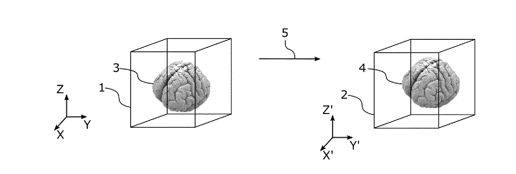
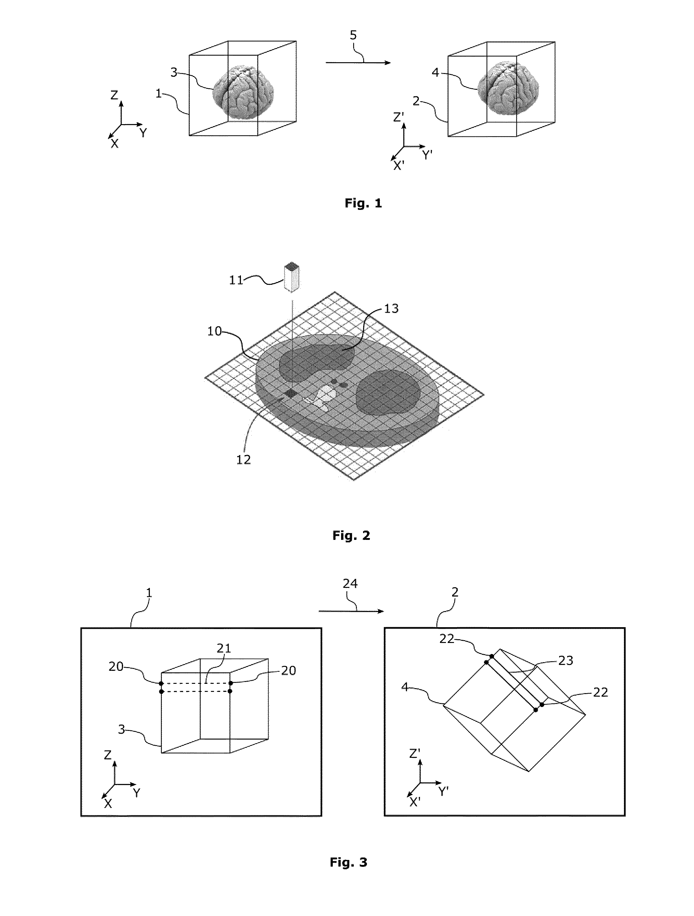
[0083]With reference to FIG. 1, the method of the invention allows calculating a region of interest (ROI) 4 in a second or a destination image 2 using an initial region of interest (ROI) 3 in a first or a source image 1. With reference to FIG. 2, the source and destination images may be 3D images composed of 3D picture elements, or voxels 11. The 3D images may usually be represented as stacks of 2D images 10 composed of 2D picture elements, or pixels 12.

[0084]A region of interest (ROI) of a 3D image 3 is an area of the image whose voxels 11 have been labeled as belonging to a specific structure, for instance during a segmentation process. In the example of FIG. 1, only a ROI 3 corresponding to a brain is shown.

[0085]If the 3D image is represented as a stack of 2D images or 2D slices, the 3D ROI appears in the 2D images 10 as a collection of labeled pixels 12 forming a 2D ROI 13.

[0086]In order to be able to compare and use the images together, the source image 1 and the destination i...


PUM


Abstract

A method is provided for calculating a region of interest in a second image using an initial region of interest in a first image. The first and second images are linked by a spatial transformation function. The method includes steps of (i) generating a first polygonal mesh enclosing the initial region of interest in the first image; (ii) computing a second polygonal mesh in the second image by applying the spatial transformation function to the first polygonal mesh, and (iii) identifying image elements within the second image, that belong to the area enclosed by the second polygonal mesh. Also provided are a computer program and a system.

Description

FIELD OF THE INVENTION[0001]The invention relates to a topology-preserving transposition method for transposing a Region of Interest (ROI) from a source image to a destination image.[0002]The field of the invention is the processing and analysis of medical images.BACKGROUND OF THE INVENTION[0003]Regions of Interest, or ROI, are used to isolate or extract areas of interest in medical images, in order to perform measurements or any other tasks on these areas.[0004]Medical images include for instance images issued from Computed Tomography (CT), Magnetic Resonance Imaging (MRI), Positron Emission Tomography (PET), or medical ultrasonography.[0005]Medical images are usually represented as collections of picture elements, such as pixels for the bi-dimensional (2D) images or voxels for the tree-dimensional (3D) or volume images. ROI can be represented as a collections of picture elements identified or labelled as belonging to, for instance, an anatomical structure (lung, blood vessel, . . ...

Claims


Application Information

Patent Timeline
IPC IPC(8): G06T7/00
CPCG06T7/0012G06T7/003G06T2207/10136G06T2207/10088G06T2207/10104G06T2207/10081A61B6/469A61B6/5235G06T7/30G06T7/337
Inventor MOULIS, MARCCADERAS DE KERLEAU, CHARLES
Owner INTRASENSE
Learn More