Endoscope system and operating method thereof

a technology of endoscope and operating method, which is applied in the field of endoscope system, can solve the problems of low color separability, deterioration of reproducibility of color, and achieve the effect of improving the color separability and the visibility of the superficial blood vessels

Active Publication Date: 2015-04-02
FUJIFILM CORP
View PDF20 Cites 8 Cited by
  • Summary
  • Abstract
  • Description
  • Claims
  • Application Information

AI Technical Summary

Benefits of technology

[0027]According to the present invention, the first and second display signals D1 and D2 are produced by the matrix operation of the signal values of the plurality types of pixels, which sense both of the first and second narrowband light.

Problems solved by technology

The primary color type imaging device is used in an endoscope system that places importance on color, because of being superior in color reproducibility, though inferior in sensitivity, to the complementary color type imaging device.
On the other hand, the complementary color type imaging device, which is superior in sensitivity and inferior in color reproducibility to the primary color type imaging device, is used in an endoscope system that places importance on sensitivity.
This causes low color separability, and a blur of the superficial blood vessels due to the influence of scattered light deteriorates the visibility of the sup

Method used


Image

  • Endoscope system and operating method thereof
  • Endoscope system and operating method thereof
  • Endoscope system and operating method thereof

Examples


first embodiment

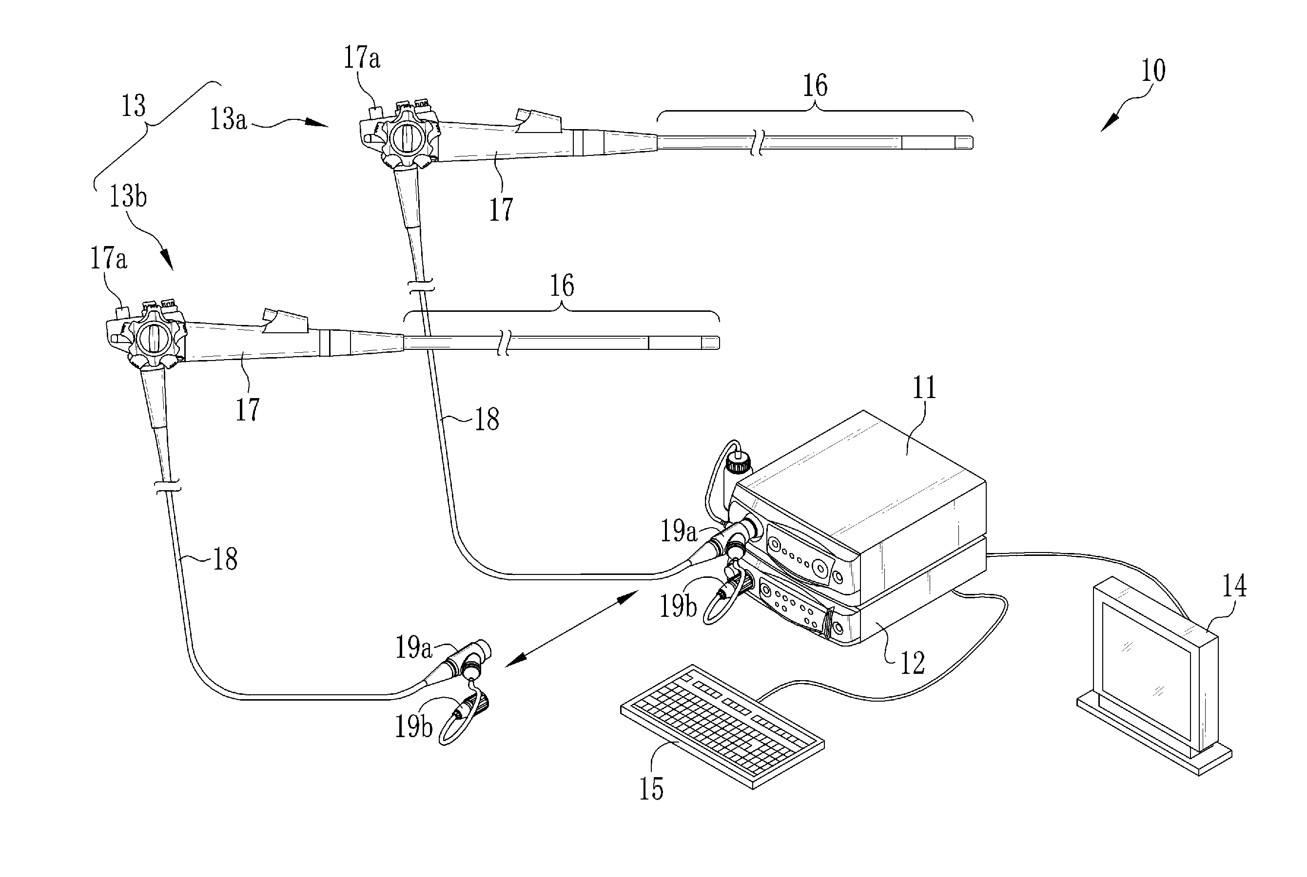
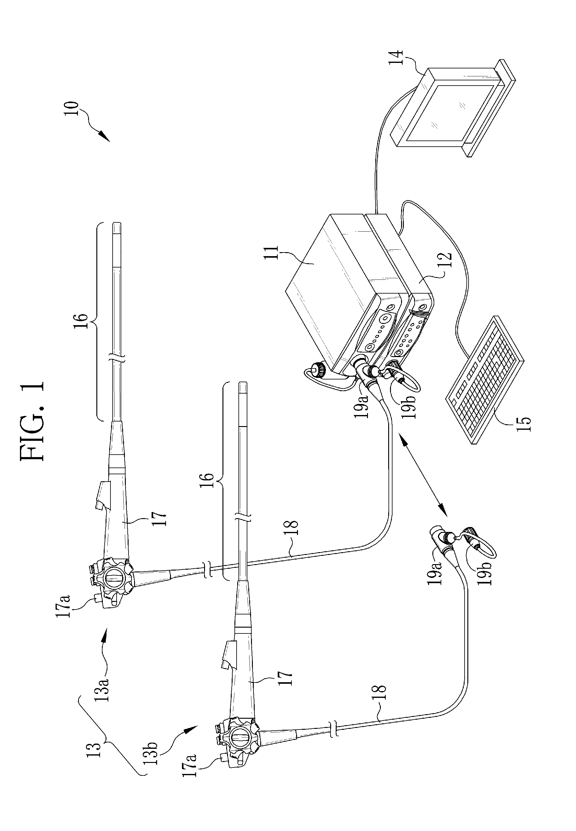
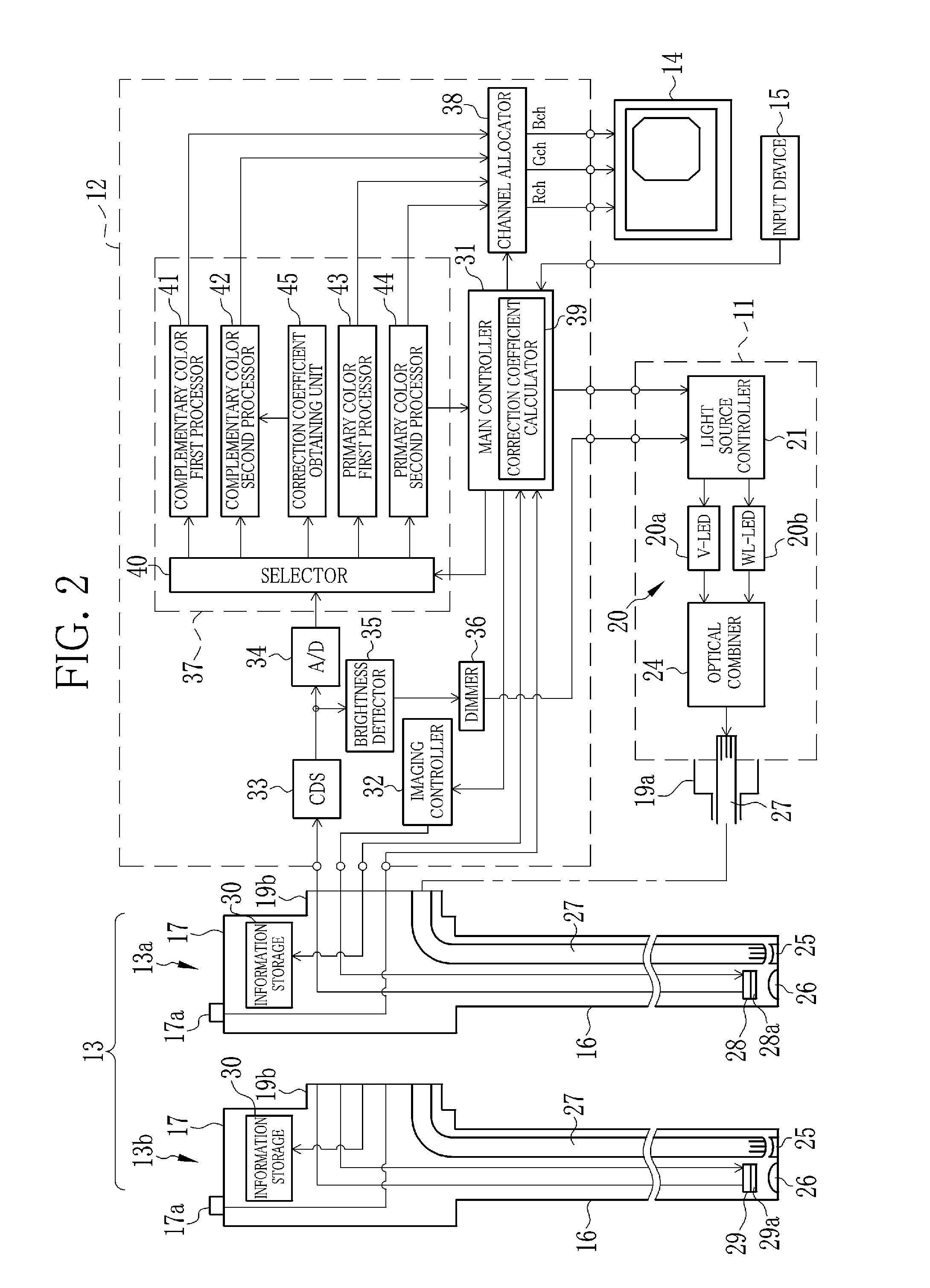
[0048]In FIG. 1, an endoscope system 10 is constituted of a light source device 11, a processor device 12, and electronic endoscopes 13 (hereinafter simply called endoscopes) detachably connected to the light source device 11 and the processor device 12. The light source device 11 produces illumination light and supplies the endoscope 13 with the illumination light. A distal end of the endoscope 13 is inserted into a human body cavity or the like to image the inside of the body cavity. The processor device 12 controls the imaging operation of the endoscope 13, and applies signal processing to an imaging signal obtained by the endoscope 13.

[0049]To the processor device 12, an image display device 14 and an input device 15 are connected. The image display device 14, being a liquid crystal display or the like, displays an image of an observation object inside the body cavity produced by the processor device 12. The input device 15, including a keyboard and a mouse, is used for inputtin...

second embodiment

[0113]Next, an endoscope system according to a second embodiment will be described. The endoscope system according to this embodiment is different from the endoscope system 10 of the first embodiment in terms that the mixed color corrector 48 performs the mixed color correction processing with the use of the following expression (10):

(D1′D2′)=(1R1R21)(1-K2-K11)(D1D2)(10)

[0114]Wherein, R1 is a first color mixture rate that represents the rate of the green narrowband light Gn component within the first display signal D1′. R2 is a second color mixture rate that represents the rate of the violet narrowband light Vn component within the second display signal D2′. R1 and R2 take values between or equal to 0 and 1.

[0115]In the case of R1=0 and R2=0, the expression (10) becomes equal to the expression (2), and the mixed color components are completely eliminated, as shown in FIG. 16. On the other hand, in the case of R1≠0 and R2≠0, the green narrowband light Gn component is present within t...


PUM


Abstract

A light source device simultaneously produces violet narrowband light and green narrowband light. A complementary color type imaging device of a complementary color type endoscope outputs first to fourth mixed pixel signals. In a complementary color second processor, a matrix operation unit performs a matrix operation of the first to fourth mixed pixel signals, and produces first and second display signals D1 and D2. A mixed color corrector corrects the first and second display signals D1 and D2 on the basis of relational expressions of “D1′=D1−K2×D2” and “D2′=D2−K1×D1”. K1 represents the ratio of a signal value of the second display signal to a signal value of the first display signal under independent application of only the violet narrowband light. K2 represents the ratio of a signal value of the first display signal to a signal value of the second display signal under independent application of only the green narrowband light.

Description

CROSS-REFERENCE TO RELATED APPLICATIONS[0001]The present application claims priority under 35 U.S.C. §119 to Japanese Patent Application No. 2013-202556 filed on Sep. 27, 2013. The above application is hereby expressly incorporated by reference, in its entirety, into the present application.BACKGROUND OF THE INVENTION[0002]1. Field of the Invention[0003]The present invention relates to an endoscope system that allows narrowband light observation and an operating method of the endoscope system.[0004]2. Description Related to the Prior Art[0005]In a recent medical field, diagnosis and treatment using an endoscope system, having a light source device, an electronic endoscope, and a processor device, are widely performed. The light source device produces illumination light and applies the illumination light to the inside of a human body cavity. The electronic endoscope images the inside of the body cavity irradiated with the illumination light by an imaging device, and produces an imagi...

Claims


Application Information

Patent Timeline
IPC IPC(8): A61B1/00A61B5/00
CPCA61B1/00096A61B5/7271A61B1/00172A61B1/00057A61B1/0638A61B1/000094
Inventor KURAMOTO, MASAYUKI
Owner FUJIFILM CORP
Why Eureka
Try Eureka