Surgery assistance apparatus, method and program

a technology of surgical assistance and equipment, applied in the field of surgical assistance equipment, method and program, can solve the problems of difficult to immediately identify and check a part of the blood vessel region, and achieve the effect of not causing damag

Active Publication Date: 2015-05-14
FUJIFILM CORP
View PDF4 Cites 11 Cited by
  • Summary
  • Abstract
  • Description
  • Claims
  • Application Information

AI Technical Summary

Benefits of technology

The surgery assistance apparatus described in this patent can help to observe blood vessels located near the cutting surface without damaging them. This is possible because the apparatus can create an image that shows the non-excision region of the cutting surface.

Problems solved by technology

The patent text discusses a technique for simulating the removal of a diseased part of an organ, such as the liver or lungs, using image-based diagnosis to determine the best way to remove the disease while preserving the function of the organ. The technique involves creating a 3D image of the organ that includes the disease, blood vessels, and other important structures. This image is then used to plan the surgery and determine which parts of the organ will be removed. The goal is to make the surgery safer and more effective.

Method used


Image

  • Surgery assistance apparatus, method and program
  • Surgery assistance apparatus, method and program
  • Surgery assistance apparatus, method and program

Examples


Embodiment Construction

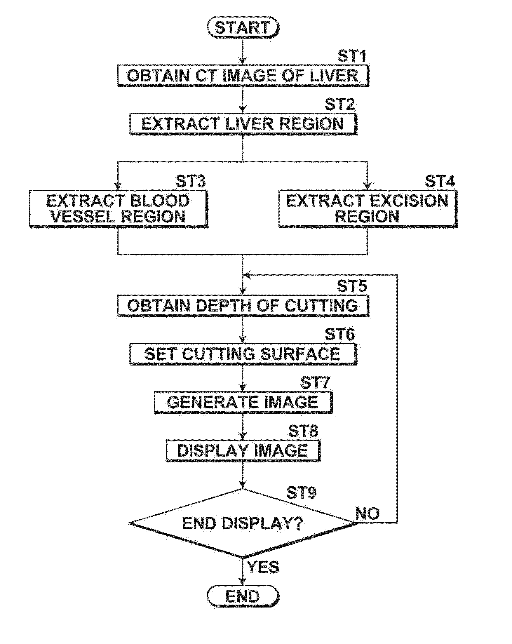
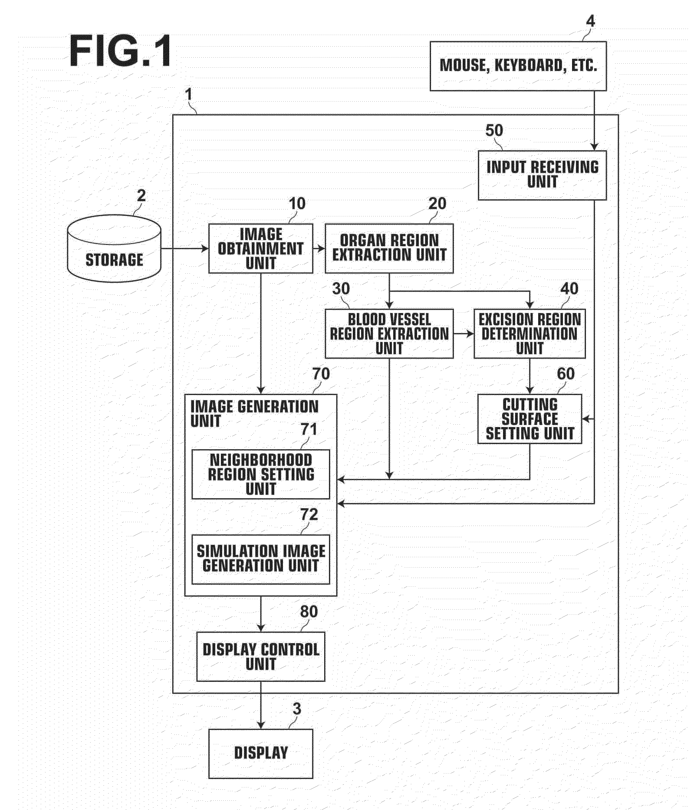
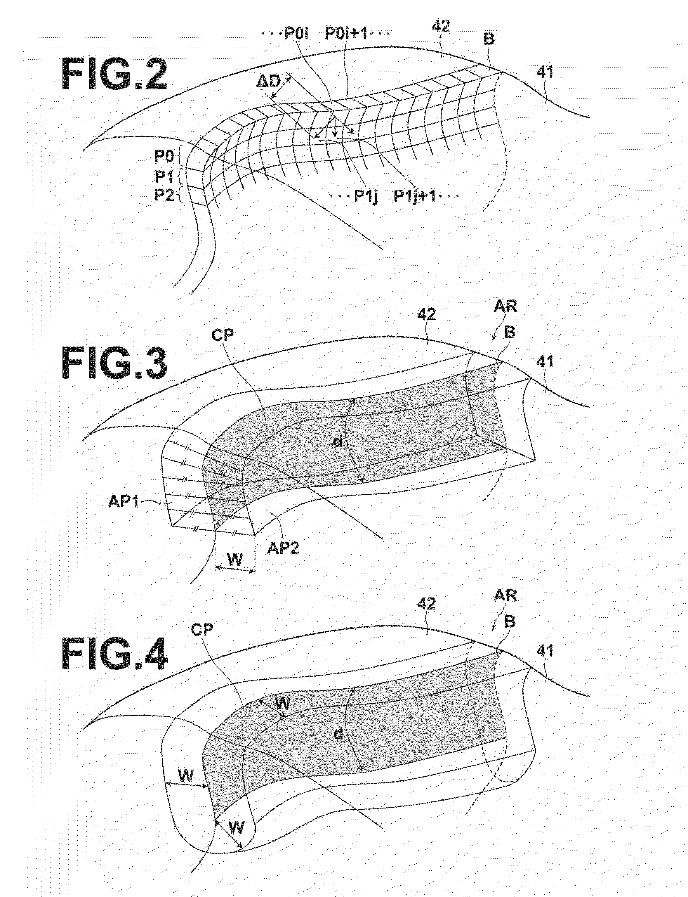
[0033]Next, an embodiment of a surgery assistance apparatus, method and program of the present invention will be described with reference to drawings. In the embodiment of the present invention, a surgery assistance system that can provide a pseudo cut-state image representing an organ, such as the liver or lungs, before or during a surgery of excising a partial region of the organ will be described. In the surgery assistance system, the set value of depth of cutting (the amount of cutting) is gradually increased as if the organ is cut gradually deeper along a surface to be cut in actual surgeries, and the pseudo-image representing the organ in a state in which the organ is cut at the depth of cutting is provided. The image represents the organ in such a manner that only partial blood vessel region VP, which is present in neighborhood region AR of cutting surface CP (a portion of the entire surface to be cut, and the portion having been cut at the depth of cutting) in blood vessel r...


PUM


Abstract

When an image representing an organ in which an excision region has been identified in such a manner that a blood vessel region in the organ is visually recognizable is generated from a three-dimensional image of the organ, an input specifying a depth of cutting is received, and a portion of a boundary surface within the specified depth of cutting along the boundary surface from an outer edge of the boundary surface toward an inside is determined as a cutting surface, and the boundary surface being between the excision region and a non-excision region in the organ. The image representing the organ in such a manner that only a partial blood vessel region, which is present in a neighborhood region of the cutting surface in the blood vessel region of the organ, is visually recognizable is generated from the three-dimensional image.

Description


Claims


Application Information

Patent Timeline
Owner FUJIFILM CORP
Why Eureka
Try Eureka