Low-dose x-ray backscatter system for three dimensional medical imaging using a conventional x-ray tube

a low-dose, x-ray technology, applied in the field of x-ray imaging, can solve the problems of loss of contrast and spatial resolution, system operation at very low exposure level, and spatial resolution of transmission x-ray system scattering of primary beam as it penetrates the breas

Inactive Publication Date: 2015-10-22
MARTIN ANNIS PATENT TRUST - 2009
View PDF0 Cites 4 Cited by
  • Summary
  • Abstract
  • Description
  • Claims
  • Application Information

AI Technical Summary

Problems solved by technology

A major limitation to the spatial resolution of transmission x-ray systems is the scattering of the primary beam as it penetrates the breast.
The fraction of scattered x-rays to the non-scattered x-rays (the only useful rays in the image) can be more than a factor of 2 for a beam of 30 keV peak at a breast thickness of approximately 5 cm, resulting in a loss of contrast and spatial resolution.
These systems operate at very low exposure levels and are limited to an exposure of 10 micro-Roentgens (μR) by government regulation.

Method used


Image

  • Low-dose x-ray backscatter system for three dimensional medical imaging using a conventional x-ray tube
  • Low-dose x-ray backscatter system for three dimensional medical imaging using a conventional x-ray tube
  • Low-dose x-ray backscatter system for three dimensional medical imaging using a conventional x-ray tube

Examples


Embodiment Construction

[0024]The present application hereby incorporates by reference in its entirety U.S. Provisional Patent Application No. 61 / 125,530, on which this application is based.

[0025]U.S. Pat. No. 7,620,150, issued to the present inventor and entitled X-ray Backscatter System for Imaging at Shallow Depths, discloses a method of producing images using a backscatter system and is incorporated herein by reference. U.S. Pat. No. 8,094,782, issued to the present inventor and entitled X-ray Backscatter System for Imaging Soft Tissue Regions, also discloses a method of producing images using a backscatter system and is incorporated herein by reference.

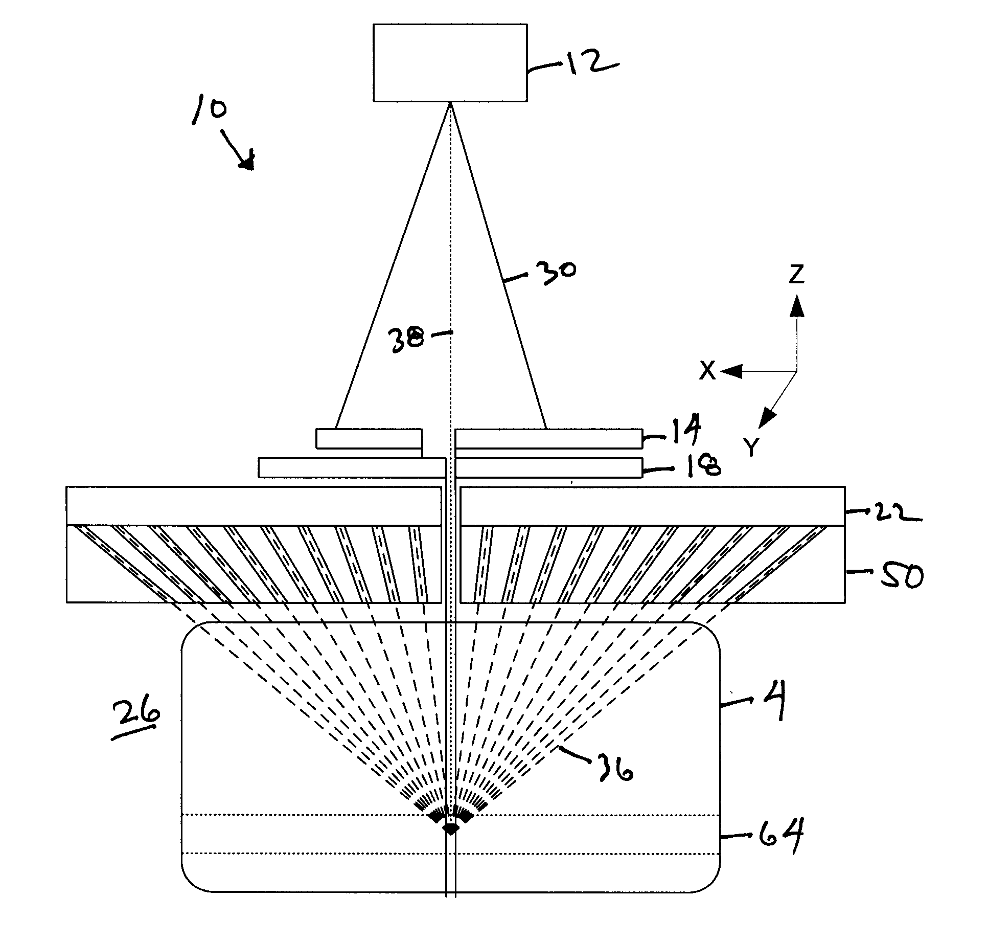
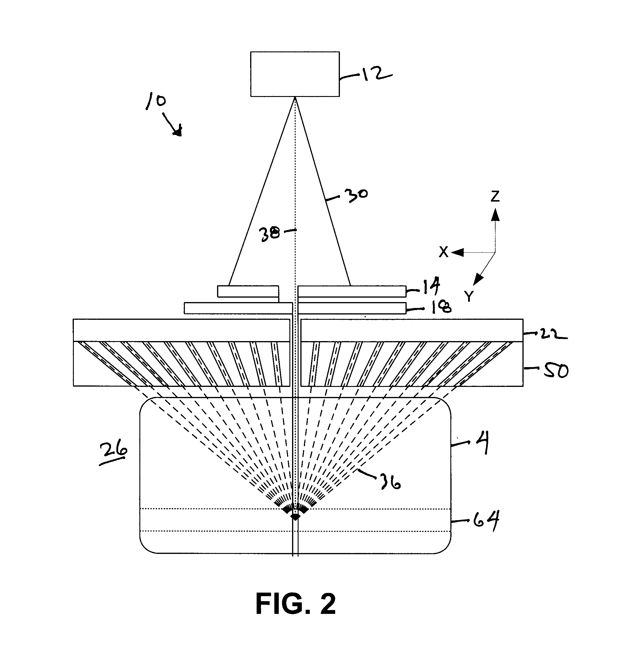
[0026]The method of the current invention is an improved version of the '782 patent and differs from the '782 patent in several ways. The '782 patent uses backscatter x-rays of the object to produce 2D images of the cancer lesion and the normal tissue simultaneously, while the current invention employs a focusing collimator that allows separate 3D imagi...


PUM

PropertyMeasurementUnit
distanceaaaaaaaaaa
distanceaaaaaaaaaa
distanceaaaaaaaaaa

Abstract

An x-ray source emits a cone beam to a rotating, x-ray-opaque disc with radial slots. The slots break the cone beam into fan beams that are emitted to an x-ray-opaque plate that produces a scanning x-ray pencil beam as each fan beam moves across a slit in the plate. A backscatter detector is adjacent to the plate. A collimator is adjacent. The pencil beam enters the object space through slits in the detector and collimator. The pencil beam moves rapidly in the y direction in the object space, producing backscatter x-rays from the object. The collimator only passes backscattered x-rays at a selected distance from the detector. Simultaneously, the assemblage of x-ray source, disc, plate, detector, and collimator moves slowly in the x and z directions. The backscattered x-rays passed by the collimator are processed to form planar images at various depths in the object space.

Description

STATEMENT REGARDING FEDERALLY SPONSORED RESEARCH OR DEVELOPMENT[0001]Not ApplicableREFERENCE TO A SEQUENCE LISTING, A TABLE, OR A COMPUTER PROGRAM LISTING COMPACT DISK APPENDIX[0002]Not ApplicableBACKGROUND OF THE INVENTION[0003]1. Field of the Invention[0004]The present invention relates to x-ray imaging, more particularly, to backscatter x-ray imaging of soft body tissue.[0005]2. Description of the Related Art[0006]X-ray imaging has been done by conventional transmission x-rays for many years. A major limitation to the spatial resolution of transmission x-ray systems is the scattering of the primary beam as it penetrates the breast. The fraction of scattered x-rays to the non-scattered x-rays (the only useful rays in the image) can be more than a factor of 2 for a beam of 30 keV peak at a breast thickness of approximately 5 cm, resulting in a loss of contrast and spatial resolution.[0007]Film imaging has now been largely replaced by digital imaging. This more convenient mode does ...

Claims


Application Information

Patent Timeline
Patent Type & Authority Applications(United States)
IPC IPC(8): A61B6/00G01N23/203A61B6/06
CPCA61B6/4071A61B6/502A61B6/06A61B6/42A61B6/4085A61B6/4078G01N23/203A61B6/0414A61B6/483
Inventor ANNIS, MARTIN
Owner MARTIN ANNIS PATENT TRUST - 2009
Why Eureka
Try Eureka