
Blood detection system with real-time capability and method of operation thereof

a detection system and real-time capability technology, applied in the field of blood detection systems, can solve the problems of small problems in surgery to become large problems, low quality of images from endoscopic or laparoscopic cameras, etc., and achieve the effects of reducing the number of surgical procedures, and improving the quality of images

Active Publication Date: 2016-01-14
SONY CORP
View PDF2 Cites 8 Cited by
  • Summary
  • Abstract
  • Description
  • Claims
  • Application Information

AI Technical Summary

Benefits of technology

The present invention is a method and system for detecting blood in an image. The system includes a camera to capture the image and a processing unit that extracts image blocks and calculates the overall blood probability of those blocks. This includes determining the red color dominance, deviation, and colorfulness. The technical effect of this invention is a more accurate and efficient way to detect blood in images, which can be useful in various fields such as medical diagnosis or security.

Problems solved by technology

However, the images from an endoscopic or laparoscopic camera can be of low quality due to a number of issues such as over- or under-exposure, insufficient light, condensation, bodily fluids obscuring the lens, or other problems.
Even with high-quality images, inattention due to fatigue or other distractions can allow small problems in surgery to become large ones.
Solutions to these problems have been long sought but prior developments have not taught or suggested any solutions and, thus, solutions to these problems have long eluded those skilled in the art.

Method used


Image

  • Blood detection system with real-time capability and method of operation thereof
  • Blood detection system with real-time capability and method of operation thereof
  • Blood detection system with real-time capability and method of operation thereof

Examples


first embodiment

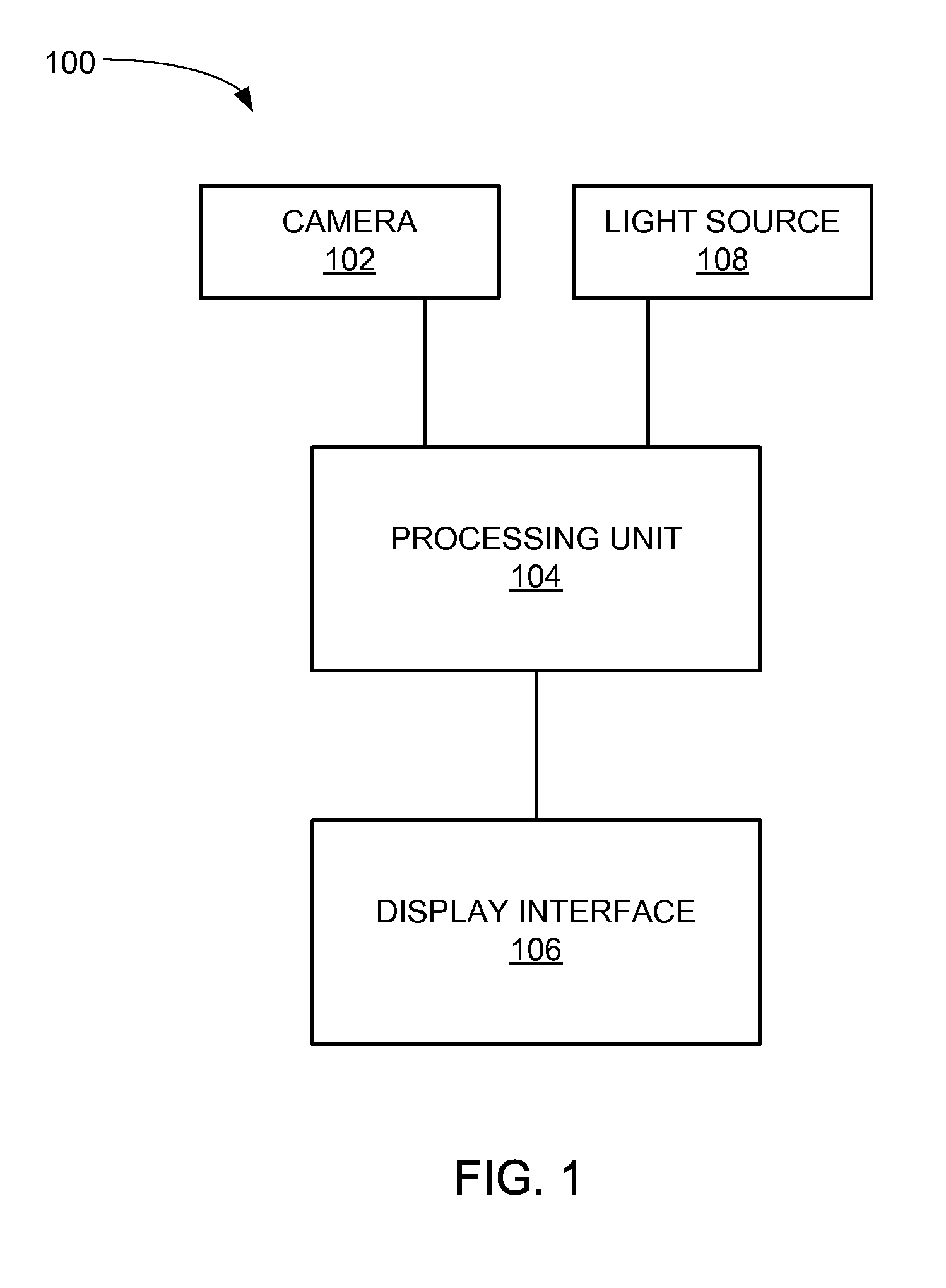
[0022]Referring now to FIG. 1, therein is shown a schematic of a blood detection system 100 in the present invention. Shown are a camera 102, a processing unit 104, and a display interface 106.

[0023]The camera 102 can be a camera capable of capturing video. The camera 102 is connected to the processing unit 104, which is connected to the display interface 106. The display interface 106 displays the view of the camera 102. Also connected to the processing unit 104 is a light source 108 for illuminating objects in view of the camera 102. The processing unit 104 is shown as connected to the light source 108 for illustrative purposes, and it is understood that the light source 108 can also be separate from the processing unit 104.

[0024]The processing unit 104 can be any of a variety of semiconductor devices such as a general purpose computer, a specialized device, embedded system, or simply a computer chip integrated with the camera and / or the display interface 106. The display interfac...

second embodiment

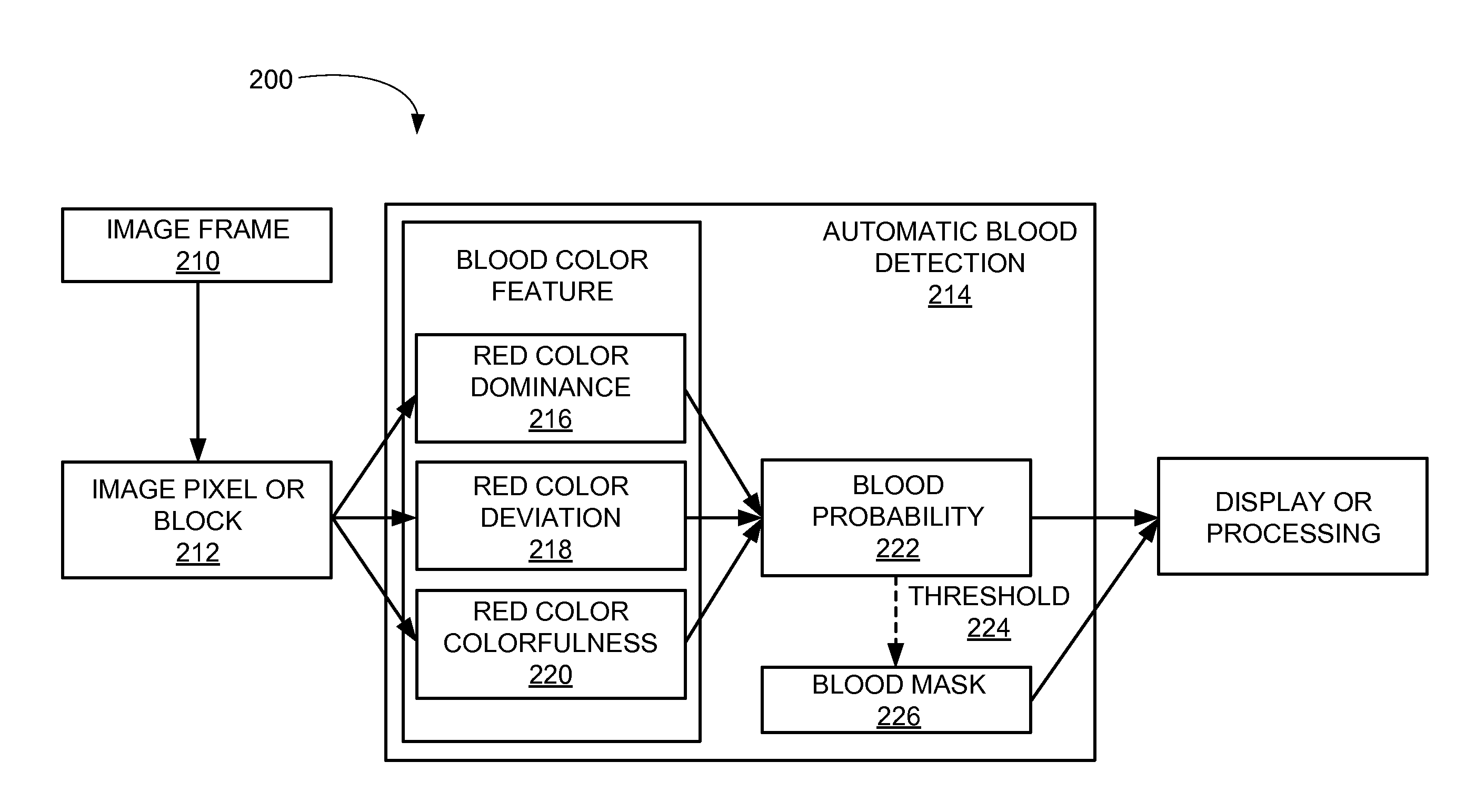
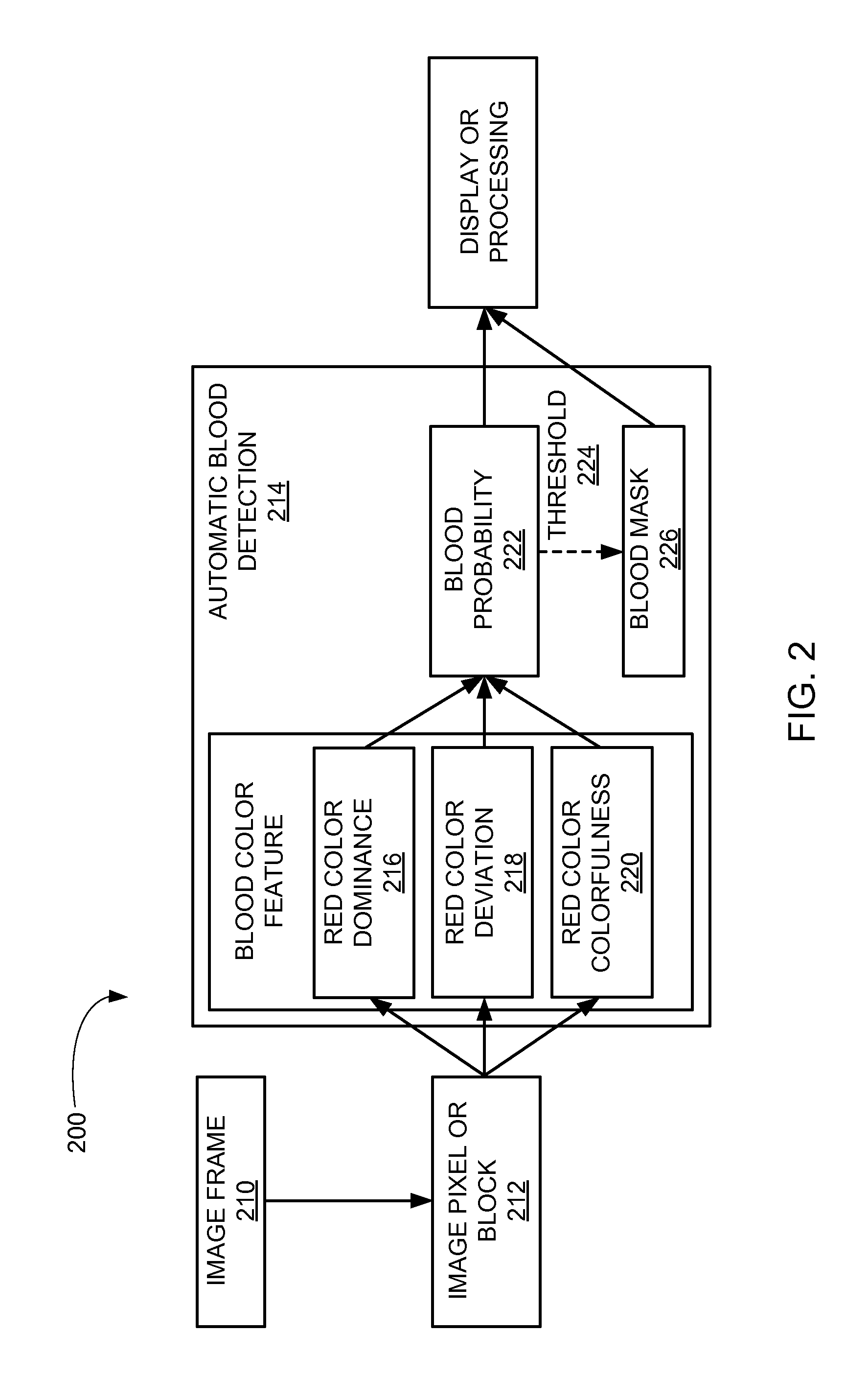
[0027]Referring now to FIG. 2, therein is shown a system diagram of the blood detection system 200 in the present invention. Starting with an input image frame 210, the image block module breaks down the input image frame 210 into image blocks 212, which can be a single pixel or groups of pixels, for example. The image blocks 212 can also be thought of as being extracted from the input image frame 210.

[0028]Each of the image blocks 212 then goes through an automatic blood detection block 214 in the automatic blood detection module. Each of the image blocks 212 goes through three separate and different blood probability tests. In this example, the feature of blood is linked to the color red. In a red color dominance test 216 the color red in each of the image blocks 212 is emphasized and a probability of blood being present in each of the image blocks 212 is calculated using the red color dominance module. In a red color deviation test 218, the deviation of the color in each of the i...


PUM


Abstract

A blood detection system, and a method of operation thereof, including: a camera for obtaining an input image frame; and a processing unit connected to the camera, the processing unit including: an image block module for extracting image blocks from the input image frame, and an automatic blood detection module, coupled to the image block module, for calculating an overall blood probability of the image blocks including: determining a red color dominance probability, determining a red color deviation probability, and determining a red color colorfulness probability.

Description

TECHNICAL FIELD[0001]The present invention relates generally to a blood detection system, and more particularly to a system for real-time detection of blood.BACKGROUND ART[0002]Advances in medical technology have improved recovery times and reduced complication rates. One significant advance is the increasing prevalence of endoscopic diagnosis and laparoscopic surgery, which avoids the need for cutting large holes in a patient by using small incisions to insert tools and a camera (i.e., endoscope or laparoscope) so the doctor or surgeon can see inside the patient. In any surgery, there are risks involved, and the ability for a physician to easily mitigate those risks is paramount to the success of the surgery.[0003]However, the images from an endoscopic or laparoscopic camera can be of low quality due to a number of issues such as over- or under-exposure, insufficient light, condensation, bodily fluids obscuring the lens, or other problems. Even with high-quality images, inattention...

Claims


Application Information

Patent Timeline
Patent Type & Authority Applications(United States)
IPC IPC(8): G06K9/46G06K9/00G06T7/00
CPCG06K9/4652G06T7/0016G06K9/00134G06T2207/30104G06K9/4642G06T2207/20076G06K9/0014G06T2207/10024G06T2207/10056G06T2207/10068G06T2207/20021G06T7/11G06T7/136G06T2207/10072
Inventor HUANG, ALBERTLIU, MING-CHANG
Owner SONY CORP
Learn More