Carbon nanotubes and graphene patches and implants for biological tissue

a technology of carbon nanotubes and graphene, applied in the field of implants, can solve the problems of recurrent herniation, affecting the safe constraint of the gel-like nucleus pulposus, and partial removal of the protruding annulus, so as to facilitate the transfer of carbon nanotubes and facilitate the attachment to adjacent tissu

Inactive Publication Date: 2016-02-04
NEW YORK SOC FOR THE RUPTURED & CRIPPLED MAINTAINING THE HOSPITAL FOR SPECIAL SURGERY
View PDF5 Cites 5 Cited by
  • Summary
  • Abstract
  • Description
  • Claims
  • Application Information

AI Technical Summary

Benefits of technology

[0011]Thus, one embodiment of the present invention is an implant or patch composition comprised of carbon nanotubes and / or graphene. The combination of the small size of carbon nanotubes and / or graphene with the extreme strength provides a patch that can be inserted into areas as small as spinal discs, but that are strong enough to withstand biological forces created by movement, such as sitting and standing. With regard to annulus fibrosus repair, this will prevent the recurrence of disc herniation and the bulge-out of the nucleus pulposus when the annulus fibrosus is damaged.
[0012]The implant or patch composition is also comprised of one or more materials that supports the nanotubes and / or graphene, and facilitates the transfer of the nanotubes to the defect. This material can be a coating. This material, which is referred to interchangeably as supportive material or supporting material or a carrier or carrier material or coating or coating material, can be any polymer, plastic, or composite material that is biocompatible. A preferred material for the supportive and / or coating material is any plastic that is hydrophobic. This material can include, but is not limited to, polyethylene glycol (PEG), polydimethylsiloxane (PDMS), vicryl, polyethylene (PE), poly-lactic acid (PLA), polyetheretherketone (PEEK), polyglycolic acid (PGA), and polyurethane. The supporting material can be modified to create binding terminals, such as amine-terminates, to create an adhesive interface that facilitates the attachment to the adjacent tissue surrounding the defect or injury. A further advantage of the patch or implant conferred by the carrier or supporting material is that the implant is flexible and has memory allowing it to be manipulated or folded when inserted or implanted in a small defect or injury in a subject and then open or unfold to its original shape and size upon insertion or implantation.

Problems solved by technology

To date, the only route to ease the pain of LDH is surgery, which holds risks for nerve damage.
While this approach provides very favorable results in the vast majority of cases (over 90% pain reduction in primary discectomy), the partial removal of the protruding annulus disturbs the safe constraint for the gel-like nucleus pulposus.
In these cases, recurrent herniations frequently occur and require repeat surgeries.
In another study, it was found that minor disc degeneration represents a risk factor for the recurrence of disc herniation after discectomy, more so than herniation volume.
However, these devices have failed as they lacked the strength to hold the herniation in place, as forces of 400 N are generated when a patient bends forward.
Therefore, novel strategies towards annular repair would significantly improve the presently limited surgical outcome in patients with contained disc herniations, but otherwise minor degenerative changes, which occur mainly in relatively young patients.
Orthopedic implants today do not allow a patient to return to normal active lifestyles, and the average lifetime of implants is only 10-15 years, meaning that younger patients endure several painful and expensive surgeries (Parchi et al.

Method used


Image

  • Carbon nanotubes and graphene patches and implants for biological tissue
  • Carbon nanotubes and graphene patches and implants for biological tissue
  • Carbon nanotubes and graphene patches and implants for biological tissue

Examples


example 1

on of an Implant Comprising Carbon Nanotubes

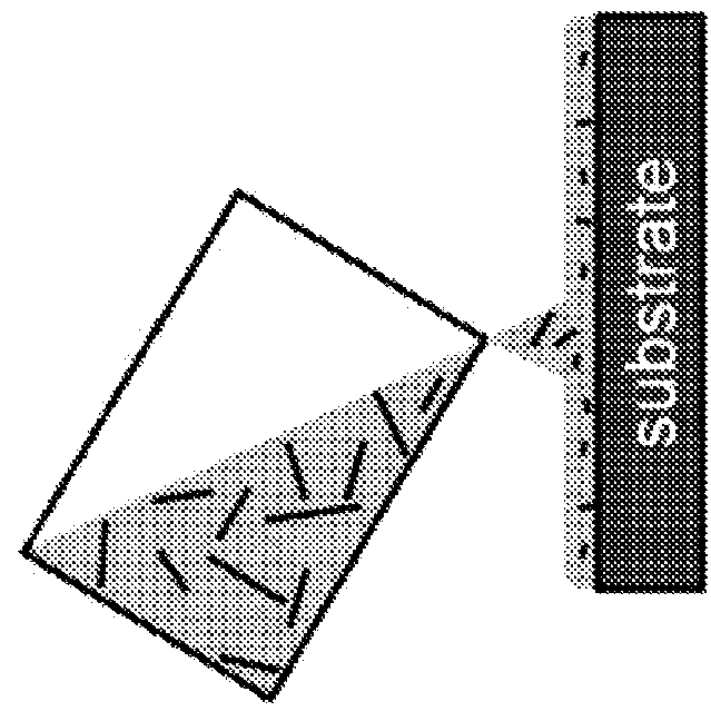
[0148]One (1) milligram of arc-discharged carbon nanotubes (Hanwha Nanotech) was dissolved in ten (10) milliliters of dichloroethane and sonicated in a bath sonicator for four (4) hours to ensure uniform dispersion of the carbon nanotubes. The solution was then centrifuged at 1000 rpm for one (1) minute to obtain a clear solution of carbon nanotubes,

[0149]The resulting carbon nanotube solution was mixed with PDMS in a 1:3 weight ratio and a PDMS hardener was added. The solution was mixed, and when thoroughly mixed was kept under vacuum for one (1) hour to remove air bubbles in solution.

[0150]The resulting mixture was poured into a substrate to form a thin film. The film was hardened on a hot plate at 60° C. for twenty (20) minutes.

[0151]A schematic of this process and the resulting patch is shown in FIG. 1.

example 2

reparation of Implants Comprising Carbon Nanotubes

[0152]Using the procedure of Example 1, implants were made using carbon nanotubes where the carbon nanotube solution was mixed with the PDMS in weight ratios ranging from 1:1.5 to 1:10, all with satisfactory results, illustrating that a range of weight ratios of carbon nanotubes to carrier can be used in the manufacture of the implants.

example 3

on of an Implant Comprising Graphene

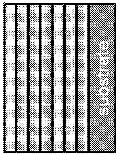
[0153]A large-scale graphene on copper foil was grown by the CVD growth method. Graphene on copper was laminated onto a thin PDMS film and copper was etched with ammonium persulfate solution.

After the copper was fully removed, the sample was gently rinsed with water and air dried.

[0154]PDMS which was pre-mixed with a hardener, was spincoated onto the graphene transferred PDMS film. The PDMS was baked at 60° C. to harden.

[0155]Another graphene on copper foil was laminated onto the film.

[0156]This process was repeated four or five times, to create a multilayer graphene patch.

[0157]A schematic of this process and the resulting patch is shown in FIG. 2.


PUM

PropertyMeasurementUnit
forcesaaaaaaaaaa
thickaaaaaaaaaa
tensile strengthaaaaaaaaaa

Abstract

The present invention relates to a novel composition comprising carbon nanotubes and / or graphene and a supporting material that can be used as an implant, patch or construct for the treatment, repair or replacement of biological tissue, including musculoskeletal tissue, fascia tissue, dura tissue, epidermal tissue, blood vessels and arteries, and organs. The present invention is also a method of manufacturing the novel composition, and a method of using the novel composition to treat, repair or replace biological tissue.

Description

CROSS-REFERENCE TO PRIOR APPLICATION[0001]The present application claims priority to U.S. patent application Ser. No. 61 / 789,384 filed Mar. 15, 2013, which is hereby incorporated by reference in its entirety.FIELD OF THE INVENTION[0002]The present invention is in the field of implants, more specifically implants or patches made of carbon nanotubes and graphene. The invention includes the implants or patches themselves, a method of making the implants or patches, and a method of using the implants or patches to repair, replace and / or treat injured and / or defective biological tissue.BACKGROUND OF THE INVENTION[0003]Lumbar disc herniation (LDH) is the painful and debilitating pathological condition for which spinal surgery is most often performed. The average age for disc herniation is 40.8 years, with ages ranging from 15-74 years (Spangfort et al. (1972)). Surgery is done most often at the level of L5 / S1 (50.5%) and L4 / 5 (47.5%). The incidence of disc surgery is growing both in the U...

Claims


Application Information

Patent Timeline
Patent Type & Authority Applications(United States)
IPC IPC(8): A61L27/44A61F2/02
CPCA61L27/443A61F2/02A61L2430/20A61L2430/34A61L2430/30A61L2400/12B82Y5/00B82Y30/00A61F2/08A61F2/28A61F2/442
Inventor SCHROEDER, JOSHUANUCKOLLS, COLINCAMMISA, JR., FRANK P.ABJORNSON, CELESTEKIM, BUMJUNG
Owner NEW YORK SOC FOR THE RUPTURED & CRIPPLED MAINTAINING THE HOSPITAL FOR SPECIAL SURGERY
Why Eureka
Try Eureka