Systems and methods for detecting tissue contact during ablation

a tissue contact and ablation technology, applied in the field of tissue contact detection systems, can solve problems such as patient serious complications, and achieve the effect of preventing ablative energy delivery

Active Publication Date: 2016-03-03
MEDTRONIC ABLATION FRONTIERS
View PDF9 Cites 55 Cited by
  • Summary
  • Abstract
  • Description
  • Claims
  • Application Information

AI Technical Summary

Benefits of technology

[0009]A medical system is disclosed, including a medical device having a plurality of electrodes; a control unit coupled to the medical device, the control unit programmed to: obtain an impedance measurement from each of the plurality of electrodes; determine whether any of the plurality of electrodes is located within the pulmonary vein based at least in part on the obtained impedance measurements; and generate an indication if at least one of the plurality of electrodes is determined to be located within the pulmonary vein. The control unit may determine whether any of the plurality of electrodes is located within the pulmonary vein by comparing an obtained impedance measurement to a predefined impedance value and / or by comparing one of the obtained impedance measurements to another one of the obtained impedance measurements. The control unit may be programmed to prevent ablative energy delivery through an electrode determined to be located within the pulmonary vein.

Problems solved by technology

However, ablation extending into the pulmonary veins can contribute to serious complications for the patient, including pulmonary vein stenosis, phrenic nerve injury, and embolic events.
However, minimally-invasive ablation procedures are typically performed within a beating heart, and the tortuous, moving cardiac environment presents challenges in accurately locating a targeted tissue and positioning a medical device in place to treat the tissue, even with external imaging systems.

Method used


Image

  • Systems and methods for detecting tissue contact during ablation
  • Systems and methods for detecting tissue contact during ablation
  • Systems and methods for detecting tissue contact during ablation

Examples


Embodiment Construction

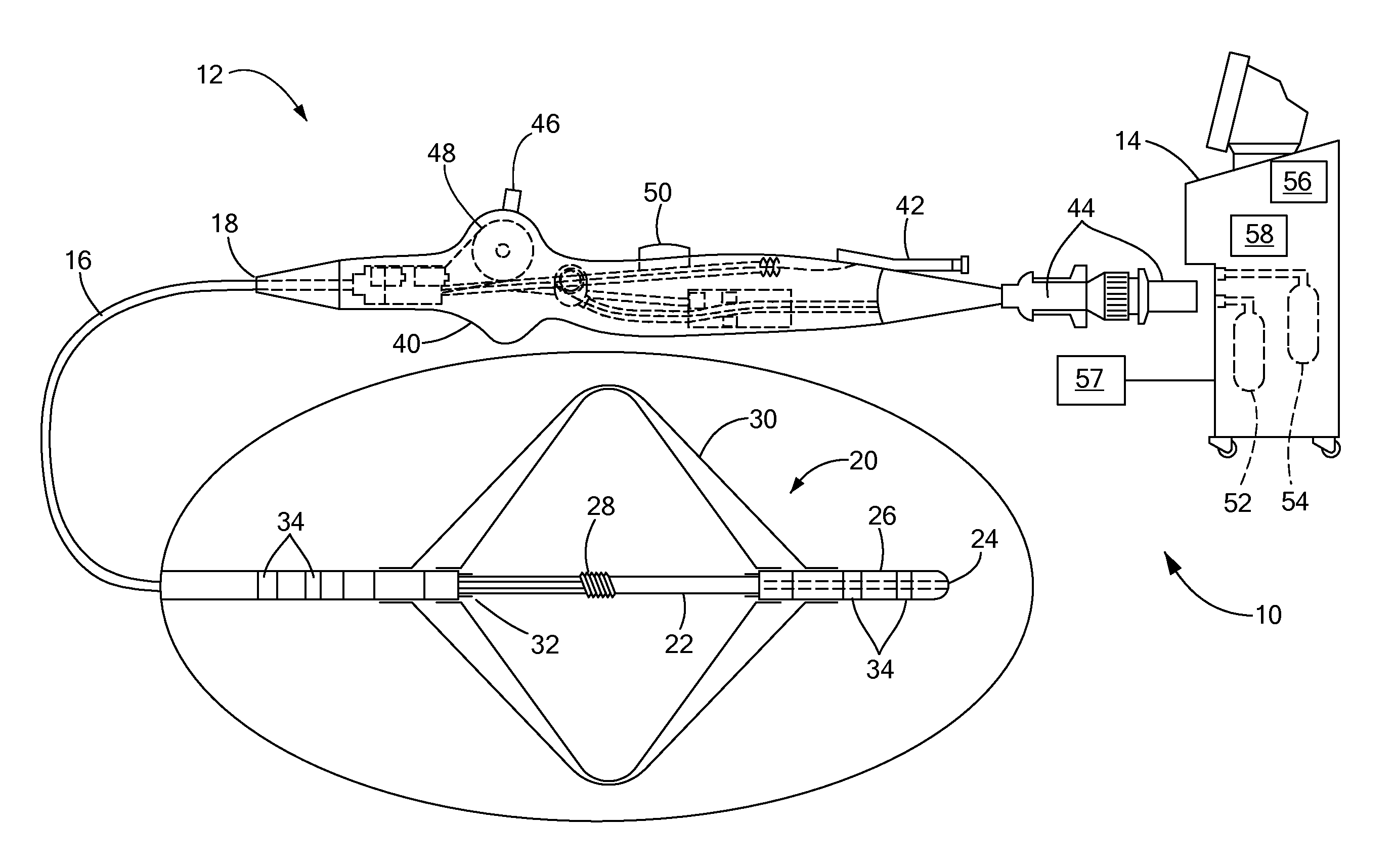
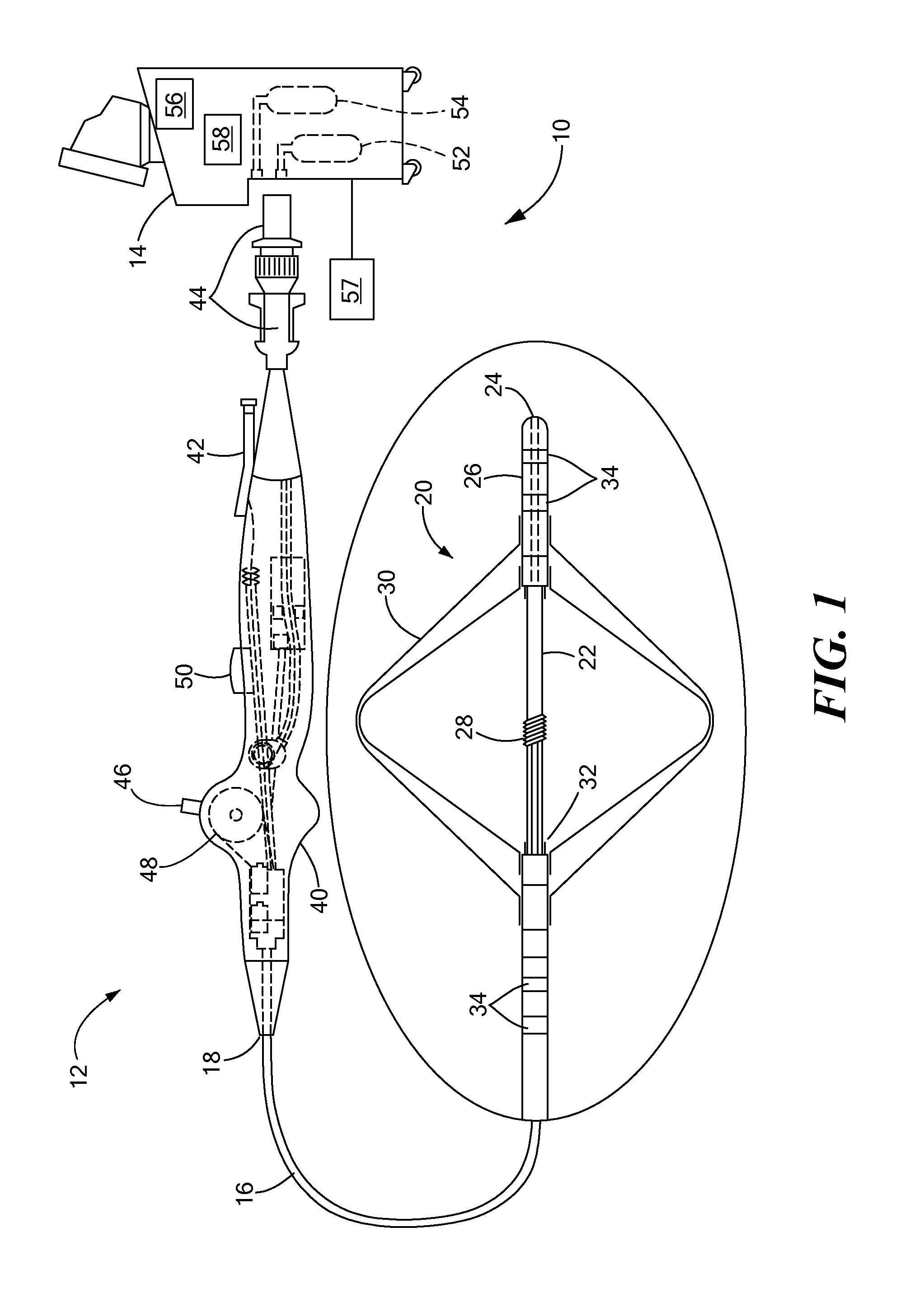
[0022]The present disclosure advantageously provides a system and methods of use thereof that facilitate accurate positioning of a medical device within a patient for subsequent treatment while avoiding damaging surrounding structures or tissue regions that are not the target of a therapeutic or diagnostic procedure. Referring now to the drawing figures in which like reference designations refer to like elements, an embodiment of a medical system constructed in accordance with principles of the present invention is shown in FIG. 1 and generally designated as “10.” The system 10 generally includes a medical device 12 that may be coupled to a control unit 14 or operating console. The medical device 12 may generally include one or more diagnostic or treatment regions for energetic, therapeutic and / or investigatory interaction between the medical device 12 and a treatment site. The treatment region(s) may deliver, for example, cryogenic therapy, radiofrequency energy, or other energetic...


PUM


Abstract

Systems and methods for assessing tissue contact, including positioning an electrode adjacent a tissue region; delivering radiofrequency energy to the electrode, wherein the delivered radiofrequency energy is sufficient to ablate at least a portion of the tissue region; obtaining a plurality of impedance measurements from the electrode during the delivery of the radiofrequency energy; calculating a change in the impedance measurements over a pre-determined time period; and generating an alert if the calculated change is less than a pre-defined value.

Description

CROSS-REFERENCE TO RELATED APPLICATION[0001]This application is a divisional of patent application Ser. No. 13 / 460900, filed May 1, 2012, entitled SYSTEMS AND METHODS FOR DETECTING TISSUE CONTACT DURING ABLATION, the entirety of which is incorporated herein by reference.STATEMENT REGARDING FEDERALLY SPONSORED RESEARCH OR DEVELOPMENT[0002]n / aFIELD OF THE INVENTION[0003]The present invention relates to a method and system for the detection of tissue contact during treatment or diagnostic procedures.BACKGROUND OF THE INVENTION[0004]Radiofrequency (RF) and cryogenic ablation procedures are well recognized treatments for vascular and cardiac diseases such as atrial fibrillation. The application of either RF or cryogenic treatment is usually based on the preference of the physician or the specific tissue to be treated. In either RF or cryogenic ablation, however, the location and quality of the lesion produced is a primary concern. The clinical success of cardiac tissue ablation to treat ...

Claims


Application Information

Patent Timeline
Patent Type & Authority Applications(United States)
IPC IPC(8): A61B18/14
CPCA61B18/1492A61B2019/465A61B2018/0022A61B2018/00285A61B2018/00345A61B2018/00577A61B2018/00642A61B2018/00708A61B2018/00791A61B2018/00863A61B2018/00875A61B2018/00898A61B2018/0212A61B2018/0262A61B2018/1467A61B19/46A61B90/06A61B2090/065
Inventor CONDIE, CATHERINE, R.SHERMAN, MARSHALL, L.STEWART, MARK, T.
Owner MEDTRONIC ABLATION FRONTIERS
Why Eureka
Try Eureka